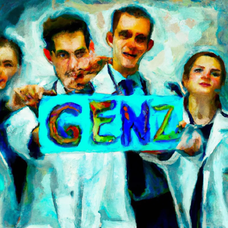
Generation Z and the younger cohort of Millennials now comprise a significant part of our working world and stir conversation, and raise questions across various fields, healthcare being no exc...
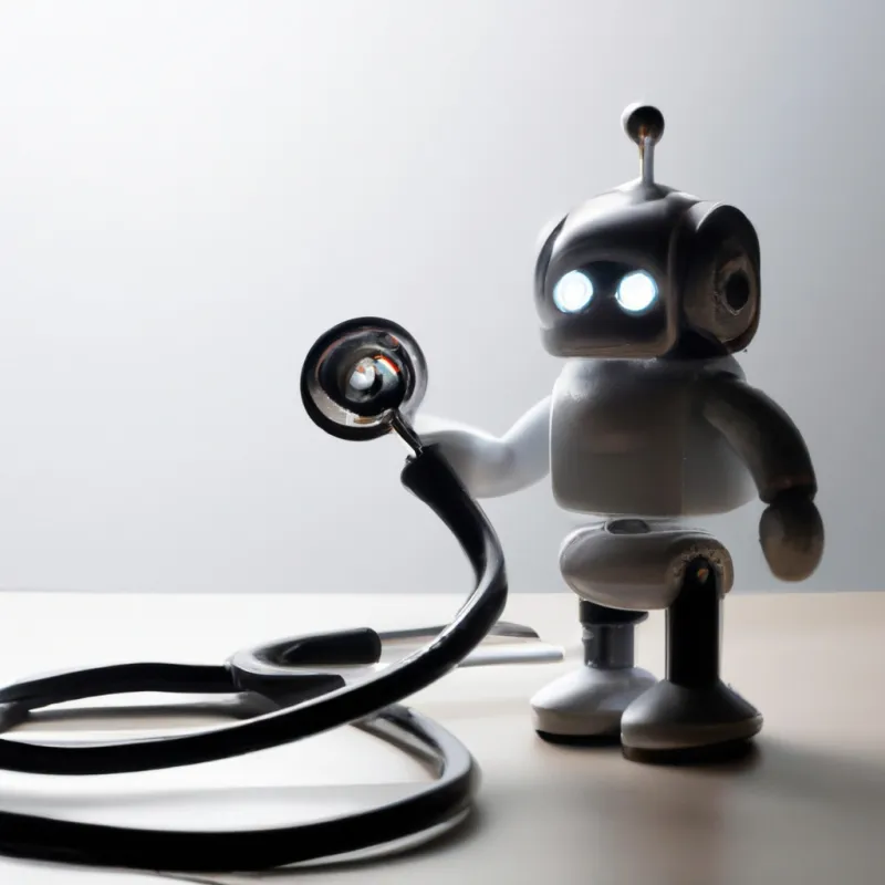
This article sums up the unique challenges that hinder complete replacement by AI while emphasizing the need to overcome these obstacles to fully harness ultrasound's potential in clinical practice.