


Course Speakers


Curriculum
Chapter 1
Free lectures
3 lectures and 0 quizzes
Lectures & Quizzes:
Chapter 2
0.25 CME
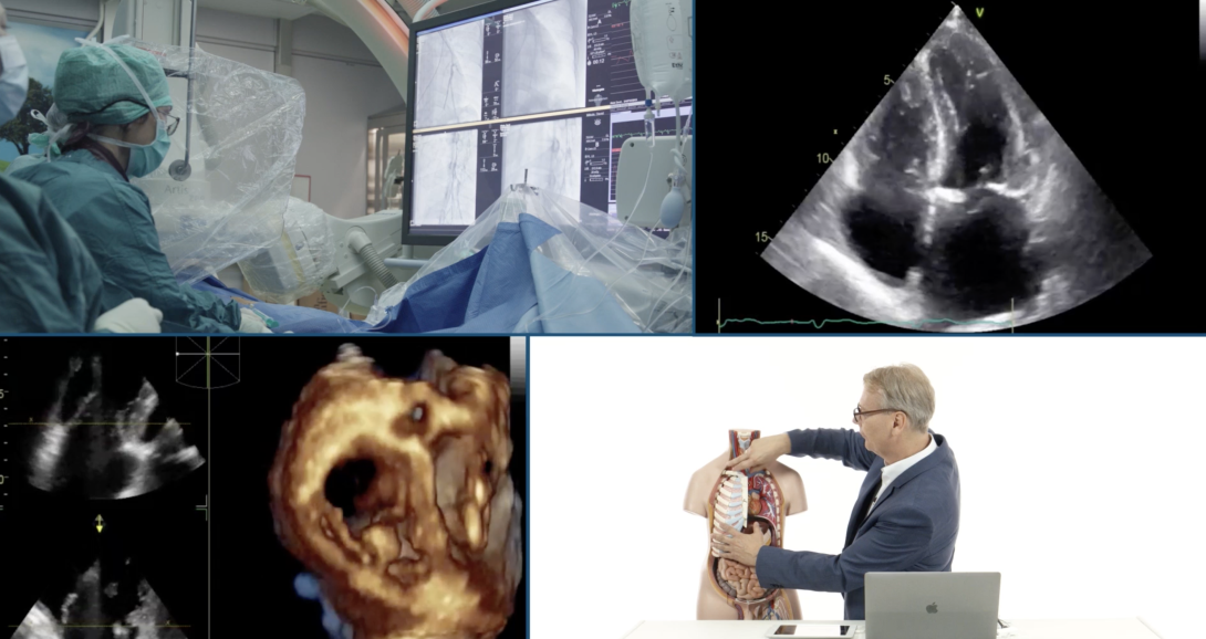
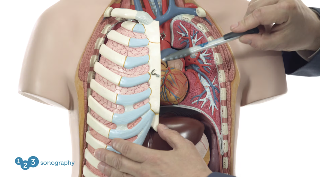
Anatomy of the Right Heart
The anatomy of the right heart is distinctly different than that of the left heart. If you want to assess right heart pathologies it is important to know the differences. In this chapter we will give you a detailed explanation of right heart anatomy and demonstrate how the specific features of the right atrium and right ventricle can be identified on the echocardiogram. Watch demonstrations on real anatomy specimens, intracardiac angioscopic videos and see lots of echo examples.
1 lectures and 1 quizzes
Lectures & Quizzes:
Chapter 3
0.25 CME
Physiology of the Right Heart
The pressures found within pulmonary circulation are significantly lower than those in systemic circulation. For this reason the right heart contracts and functions differently than the left. In this chapter, we will cover right heart physiology and discuss what happens if the right heart is confronted with an increase or decrease in afterload.
1 lectures and 1 quizzes
Lectures & Quizzes:
Chapter 4
0.25 CME
Echocardiographic Assessment - How to Image the Right Heart
Imaging of the right heart using standard views is an essential part of every echo exam. But if you want to assess right heart pathologies, it is often necessary to use additional and sometimes even "exotic" views. In this chapter, we will show you how you can get more out of your exam. Here you will see many live imaging demonstrations, simulations and echo examples.
1 lectures and 1 quizzes
Lectures & Quizzes:
Chapter 5
0.25 CME
Echocardiographic Assessment - Size
Dilatation of the right heart is a common feature of right heart disease. But how can you tell if the right heart is truly dilated? What are the normal values? What is the degree of right heart enlargement? In this chapter, we will focus on the various measurements proposed by the guidelines, provide you with the normal values, and discuss the advantages and disadvantages of the individual parameters.
1 lectures and 1 quizzes
Lectures & Quizzes:
Chapter 6
0.5 CME
Echocardiographic Assessment - Function
Quantification of right heart function is quite tricky and more difficult than the quantification of left heart function. There are several different parameters that can be used. These include "eyeballing", TAPSE, fractional area change, tissue Doppler of the RV, and right ventricular strain. Here we will show you how to obtain these individual measurements and how they can be used in an "integrative approach" to quantifying right heart function.
1 lectures and 1 quizzes
Lectures & Quizzes:
Chapter 7
0.25 CME
Echocardiographic Assessment - Pulmonary Pressure
The ability of echocardiography to quantify pulmonary pressures is one of the main reasons why this method is so important in right heart disease. But how is it done correctly? What do the values mean? In which clinical scenarios is it important to know how high pulmonary pressure is? What do we do if there is no adequate TR signal from which we can calculate pulmonary pressures? These and many other questions will be answered here.
1 lectures and 1 quizzes
Lectures & Quizzes:
Chapter 8
0.5 CME
Role of Radiology - Part 1: Overview, Chest X-Ray, CT, Pulmonary Angiography
Radiology plays an important role in the assessment of right heart disease. Florian Wolf, an expert in the field of cardiovascular imaging, will guide you through the wide spectrum of modalities that radiology offers. In this chapter, we will give you an overview of the different modalities and then focus on CT, Angio-CT, and Pulmonary Angiography. Learn how they are performed, when they are applied, and what the risks are.
1 lectures and 1 quizzes
Lectures & Quizzes:
Chapter 9
0.25 CME
Role of Radiology - Part 2: MRI, Clinical Examples in Radiology
In the second part dealing with Radiology, Florian Wolf will focus on the role of magnetic resonance imaging and show you several cases of patients with a right heart pathology and how CT and MRI can help to detect the problem.
1 lectures and 1 quizzes
Lectures & Quizzes:
Chapter 10
0.5 CME
Pulmonary Hypertension - Classification/Pathophysiology
Pulmonary hypertension can have many causes. The international societies propose a classification scheme that guides us through the 'jungle' of different disease entities. In this chapter, Irene Lang, a leading expert in the field of pulmonary hypertension, will explain the pathophysiology of PHT and discuss the different causes of pulmonary hypertension.
1 lectures and 1 quizzes
Lectures & Quizzes:
Chapter 11
0.5 CME
Pulmonary Hypertension - Diagnosis: Overview Part 1 (+ Right Heart Cath)
Signs and symptoms of pulmonary hypertension can be quite vague and unspecific. It is not uncommon that there is a long delay until the diagnosis is established. In this chapter, we introduce you to a patient with pulmonary hypertension who will share her personal story with you. She will tell you how she was diagnosed and how the disease impacts her life. In addition, we will discuss the individual diagnostic tests that are applied in the workup of patients inclduing: BNP, 6 min walk test, CT scan, and most importantly, right heart catheter. Here we will take you to the cath lab to show you a complete procedure.
1 lectures and 1 quizzes
Lectures & Quizzes:
Chapter 12
0.25 CME
Pulmonary Hypertension - Diagnosis: Overview Part 2
In the second part of this course dealing with the diagnosis of pulmonary hypertension, we will discuss issues including as the role of the ECG, the six minute walk test, and pulmonary function testing. Here we will also show you the typical features of CTEPH and PAH on the pulmonary angiogram.
1 lectures and 1 quizzes
Lectures & Quizzes:
Chapter 13
0.25 CME
Pulmonary Hypertension - Diagnosis: Echo Features of PHT
Echocardiography plays an important role in pulmonary hypertension — not only in the screening of patients, but also in assessing prognosis and monitoring of treatment effects. This chapter will provide you with the typical echo features of pulmonary hypertension such as right ventricular enlargement, the "D-shaped" ventricle, right ventricular dysfunction, poor cardiac output, and more. You will learn how these findings impact prognosis and how they are used in various clinical scenarios.
1 lectures and 1 quizzes
Lectures & Quizzes:
Chapter 14
0.5 CME
Pulmonary Hypertension - Therapy: Drugs
Tremendous achievements have been made in the treatment of pulmonary hypertension and we now have numerous, highly effective drugs at hand. In his chapter, we explain how medication alters prognosis, which class of drugs are available, how to use a risk score to guide medication, and why combination therapy is important.
1 lectures and 1 quizzes
Lectures & Quizzes:
Chapter 15
0.25 CME
Pulmonary Hypertension - Interventional and Surgical Techniques
Interventional and surgical techniques such as Balloon Pulmonary Angioplasty, Thromboendarterectomy, and Lung Transplantation are important options for patients with pulmonary hypertension. But which patients are potential candidates for these therapies? How are they performed? And what is their impact on prognosis? All of these questions will be answered in this chapter.
1 lectures and 1 quizzes
Lectures & Quizzes:
Chapter 16
0.5 CME
Right Heart Volume Overload
Right ventricular volume overload can have disastrous effects on the right heart. But when does it occur? How does it affect the right heart? Which abnormalities do we see on the echo and what is its consequence? These topics will be discussed here. Since tricuspid regurgitation (TR) is the most common cause of right ventricular volume overload, we will also focus on the tricuspid valve and its pathologies. You will also learn how to quantify TR and which treatment options for TR are available. Other topics related to volume overload covered in this chapter are pulmonic regurgitation and atrial septal defects.
1 lectures and 1 quizzes
Lectures & Quizzes:
Chapter 17
0.5 CME
Pulmonary Embolism
Watch this chapter to better understand pulmonary embolism (PE) and its sequels. Here you will learn when to suspect PE, how to diagnose it, which pathologies can mimic PE, what impacts prognosis and how echocardiography can help you to stratify patients into high and low risk groups. We will present several cases that will demonstrate the typical echocardiographic features of acute pressure overload.
1 lectures and 1 quizzes
Lectures & Quizzes:
Chapter 18
0.5 CME
The Right Heart in CAD, CMP and Valvular HD
The right heart is often affected in disease of the left heart. Left heart disease is actually the most common reason for pulmonary hypertension. Did you know that myocardial infarctions can also involve the right heart? And that right ventricular dysfunction is frequently present in patients with myocarditis and dilated and restrictive cardiomyopathy? Using a case based approach, we will cover these topics in this chapter and discuss the importance of the right ventricle is various "left heart" scenarios.
1 lectures and 1 quizzes
Lectures & Quizzes:
Chapter 19
0.25 CME
Arrhythmogenic Right Ventricular Dysplasia/Carcinoid Heart Disease
Echocardiography plays an important role in the diagnosis and management of right ventricular dysplasia and carcinoid heart disease. In both disease entities, it is important to establish the diagnosis as early as possible. In this chapter, we will show you the typical echocardiographic features of carcinoid heart disease and right ventricular dysplasia that will make it easy for you to make the diagnosis. With the help of several case examples, you will be able to put everything you have learned into a clinical context.
1 lectures and 1 quizzes
Lectures & Quizzes:
Chapter 20
0.5 CME
Right Heart in Congenital Heart Disease - Part 1
The right heart is frequently involved in congenital heart disease. In this chapter, we will provide an overview of different congenital defects that can cause volume overload, such as primum and secundum atrial septal defects, sinus venosus defects, pulmonary regurgitation / Fallot tetralogy. We will also provide clues that will help you to identify the right ventricle in complex situations.
1 lectures and 1 quizzes
Lectures & Quizzes:
Chapter 21
0.25 CME
Right Heart in Congenital Heart Disease - Part 2
This is the second chapter that deals with right heart disease in congenital heart defects. Here we will cover defects that cause pulmonary hypertension as they can occur in congenital left heart disease (post-capillary pulmonary hypertension), shunt lesions and preoperatively. The following defects will be discussed: Coarctation of the Aorta (CoA), ASD with pulmonary hypertension, ventricular septal defects, Patent ductus arteriosus (PDA), and single ventricles. Special emphasis is placed on the Eisenmenger syndrome to help you understand the pathophysiology behind, when it occurs, how patients with Eisenmenger present and which factors can lead to clinical deterioration.
1 lectures and 1 quizzes
Lectures & Quizzes:
Objectives

After completing the Right Heart MasterClass, you will know how to identify right heart diseases.
You will be able to classify the pathology and assess its prognosis.
Know how to possess a thorough understanding of the individual treatment options.
Ideal for:

Student Discount
Are you a student? Get 50% discount on this course by completing the student application form.
Get Student DiscountRecommended Blog Posts

Pricing
One-Month Access
Take the most flexible route with a monthly subscription.
You get:
- Cancellation possible anytime after 4 months minimum run time
- Ability to complete quizzes and earn CME credits
Two-Year Access
If you want to take your time learning, this option is perfect for you. Save 40% on the 6-month option.
You get:
- 24 months access to our course
- Ability to complete quizzes and earn CME credits
One-Year Access
Our most recommended access duration to dive deep into the course. Save 30% on the 6-month option.
You get:
- 12 months access to our course
- Ability to complete quizzes and earn CME credits
Half-Year Access
Our shortest option for very fast learners. Ideal for people who have plenty of time to learn.
You get:
- 6 months access to our course
- Ability to complete quizzes and earn CME credits