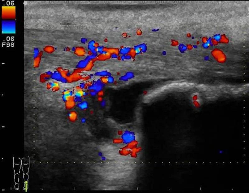
Images showing increased vascularity, excess fluid within the bursa, and soft tissue swelling indicating the presence of inflammation, bursitis, and achilles tendon tendinosis are discussed, along with other marke...
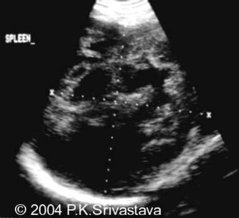
A large hypoechoic complex mass is visualized within the spleen on both ultrasound and CT warranting a biopsy which proved it to be a lymphomatous infiltration secondary to Hodgkin's lymphoma.
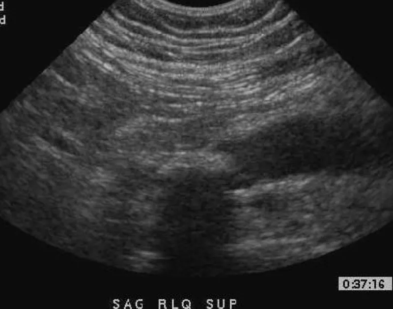
Imaging of the right lower quaderant is provided showing a non-compressable hypoechoic structure with hyperemia and calcification and the diagnosis of acute appendicitis is discussed as well as the process of diag...
Along with a discussion of other possible locations and presentations of metastatic lymph adenopathy, images depicting abdominal metastatic adenopathy and associated ascites are presented in the setting of diffuse...
Imaging from a woman with acute right upper quadrant pain is shown, illustrating a thickened gallbladder wall, cholelithiasis, hyperemia, and pericholecystic fluid, constant with acute cholecystitis.
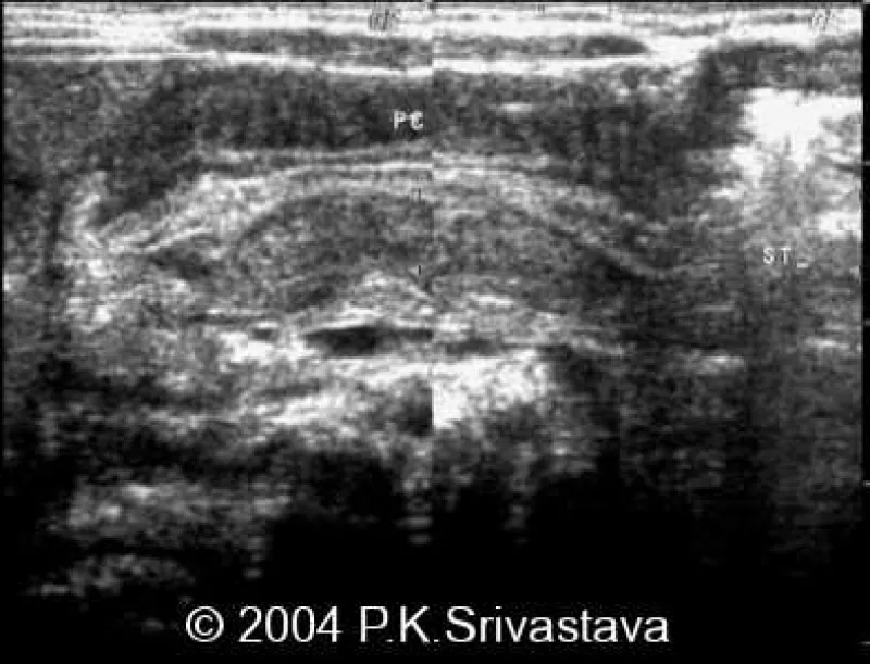
The condition of pyloric stenosis is discussed, with a presentaton of a case showing an enlarged pyloris with thickened hypoechoic mucosal layers visualized.