Find the answer to our question "What is meant by the “bat sign” in ultrasound?"
Case stories that stick

What is meant by the “bat sign” in ultrasound?
Echocardiography primarily zeroes in on the heart, but did you know it can also detect non-cardiac anomalies like hepatic cysts?
With the help of M-Mode it is possible to make interesting teaching points.
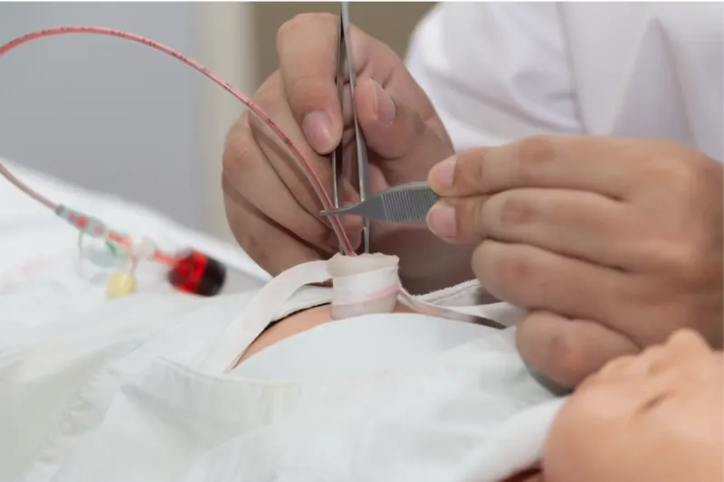
In this post, we'll delve into the importance of using Point-of-Care Ultrasound (POCUS) in the Neonatal Intensive Care Unit (NICU) by presenting a real patient case who needed umbilical vein catheterization.

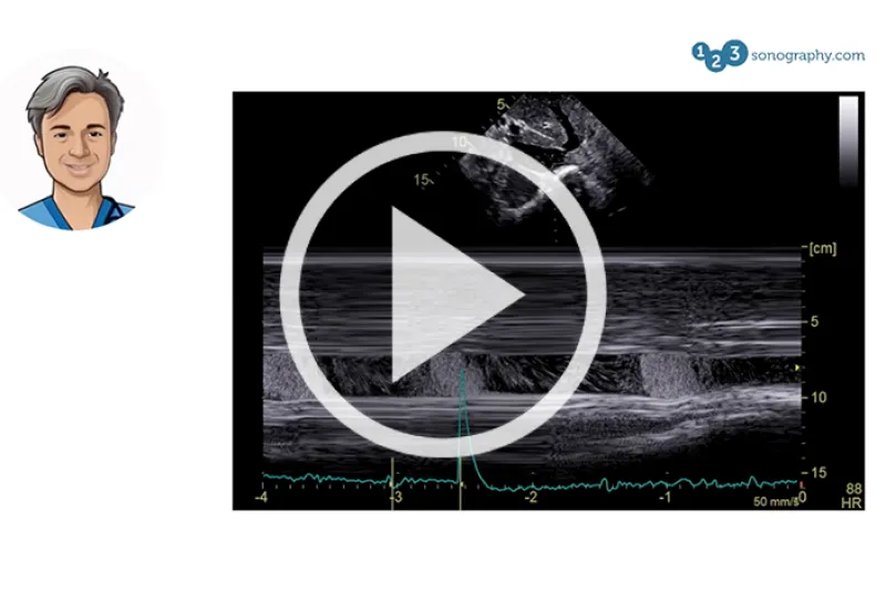
In this part, we have prepared a short video for you about documentation and quantification in ultrasound because, after all, measuring and saving our