
2.3 Two Dimensional Imaging / The Windows
The heart is surrounded by lung tissue and is hidden behind the rib cage. Both lung tissue (air!) and bone are barriers for ultrasound. This makes echocardiography more difficult than other types of ultrasound investigation, since we only have rather small windows from which the heart can be visualized.
Basically there are three principal windows to the heart: the left parasternal, the apical, and the subcostal window. Under certain circumstances we may use additional windows such as the right parasternal or the suprasternal window. Be creative: in certain settings you may use atypical windows (e.g. imaging from the back in the presence of a pleural effusion).
Imaging windows Parasternal - 2nd - 4th intercostal space left margin of the sternal boarder Apical 4th - 5th intercostal space, lateral! Subcostal Below xiphoid Right parasternal 2nd - 4th intercostal space Suprasternal Jugulum sternum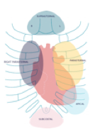
The exact location of the windows may differ from one patient to the next; not all of them can be visualized from every window. The patient's constitution, the position of the heart, and the surrounding tissue greatly influence the use and location of the imaging windows.
For example, in asthenic patients the heart may be hidden behind the sternum. This makes it difficult to image these patients from a parasternal approach. In patients with emphysema or COPD it may be difficult to obtain images from the apical window because lung tissue obstructs the investigator's view.
Situations in which it is difficult to image Very obese Abnormalities of the chest (pectus excavatum) COPD Breast implants Very slim Lung resection Constrictive pericarditisFrom each window we acquire several so-called standard views, which jointly make up the cornerstones of a complete evaluation. However, a thorough exam will require variations of these views, for which you focus on specific structures and pathologies. The sequence of windows and views is standardized. A standard exam always begins with the parasternal window and the parasternal long axis.