
2.3.4 Suprasternal Window


The suprasternal window is located in the jugulum right on top of the sternum (suprasternal notch). We do not use this window on a regular basis or in every exam, but it can be quite useful for specific situations, such as measuring the width of the aortic arch, looking for aortic dissection or coarctation, assessing retrograde flow in the descending aorta (using color Doppler) or when quantifying aortic regurgitation.
The patient should be positioned in a supine position with his/her neck extended as far as possible - ideally, the patient's head "hangs" over the edge of the bed, you can also put a pillow under the patient’s shoulders to facilitate their posture. In general, it is easier to image young individuals, because they are able to extend their head much further than older patients. Patients with emphysema (e.g. due to COPD) are difficult to image because the air in the lungs obstructs your view, which can be countered by asking them to exhale during the imaging procedure.
The ascending aorta and the aortic arch bend from the heart’s right ventral to its left caudal side. To transect the aorta in a longitudinal view you best place your index finger on the marker and align it with the orientation of the aortic arch - meaning the marker points somewhere in the direction of the patient’s left acromion or scapula. You need an oblique angle in relation to the frontal plane of the body.
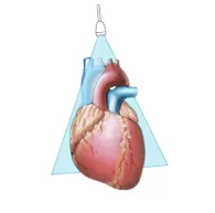
Cut plane of the suprasternal view

Suprasternal View
The suprasternal long axis shows the ascending aorta (on the left of the image), the aortic arch with the brachiocephalic artery (common trunk), the left common carotid artery, the left subclavian artery and the cranial segment of the descending aorta (on the right). The right pulmonary artery is seen in an orthogonal view as it crosses beneath the aortic arch.
You can also obtain short-axis views of the aorta by rotating the transducer 90 degrees clockwise. You will then image the pulmonary artery in the long axis and by tilting the transducer anteriorly (the transducer will almost touch the patient’s throat), you can see a structure anterior to the aorta, the innominate vein. Tilting even further inferiorly, you might be able to see the left atrium and the pulmonary veins entering it. This view is called “crab view” and can be helpful for viewing the left atrial anatomy or when you want to image the veinous inflow into the left heart.
As already mentioned, the suprasternal view allows you to look for aortic aneurysms or dissection of the aorta, so you should perform this view whenever you find a dilated aorta in a parasternal view. Intima flaps can often be seen nicely from this perspective, allowing you to spot problematic alterations of the aorta early and easily.
Specific uses of the suprasternal view
- Measure the width of the aortic arch
- Look for aortic dissection or coarctation
- Assess retrograde flow in the descending aorta