Introduction to the Subcostal View - lecture for beginners.
Case stories that stick
Introduction to the Apical Window - lecture for beginners.
You will learn how to hold the transducer, how to position, where to put the transducer and how to manipulate it in order to find the optimal position.
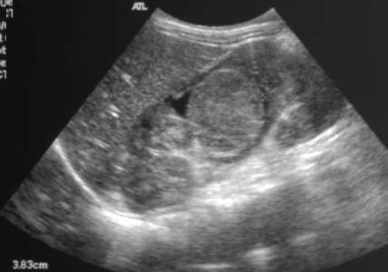
Both ultrasound and CT images are presented illustrating the presence of wilms tumor and the extent of the disease process, and the incidence, histology, and symptoms of this highly curable pediatric condition are...
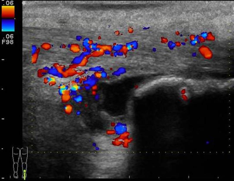
Images showing increased vascularity, excess fluid within the bursa, and soft tissue swelling indicating the presence of inflammation, bursitis, and achilles tendon tendinosis are discussed, along with other marke...
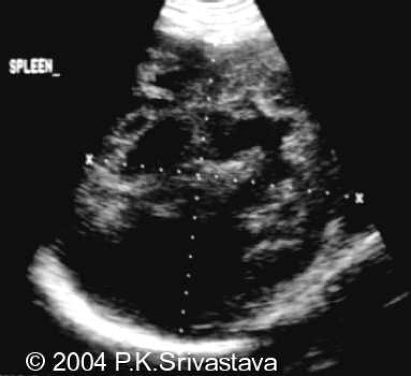
A large hypoechoic complex mass is visualized within the spleen on both ultrasound and CT warranting a biopsy which proved it to be a lymphomatous infiltration secondary to Hodgkin's lymphoma.


