
Retrocalcaneal bursitis and Achilles tendon tendinosis

First published on SonoWorld
Case Presentation
30 year old athlete with persistent heel pain on the right side
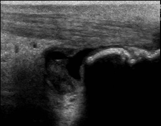 Caption: Longitudinal image of the right distal Achilles tendon | Description: There is a fluid filled thick walled bursa dorsal of the achilles tendon
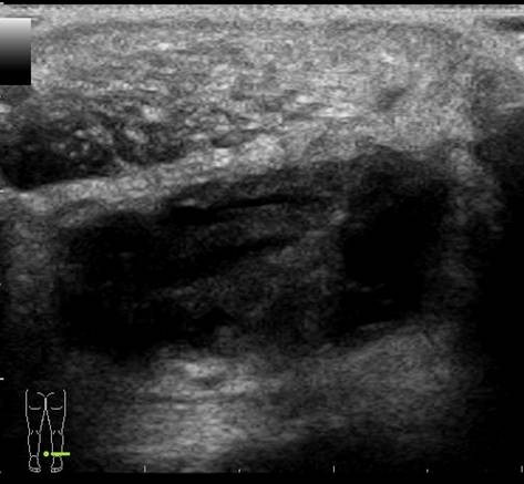 Caption: Transverse image of the right distal Achilles tendon | Description: There is a fluid filled thick walled bursa dorsal of the achilles tendon with rounded contours
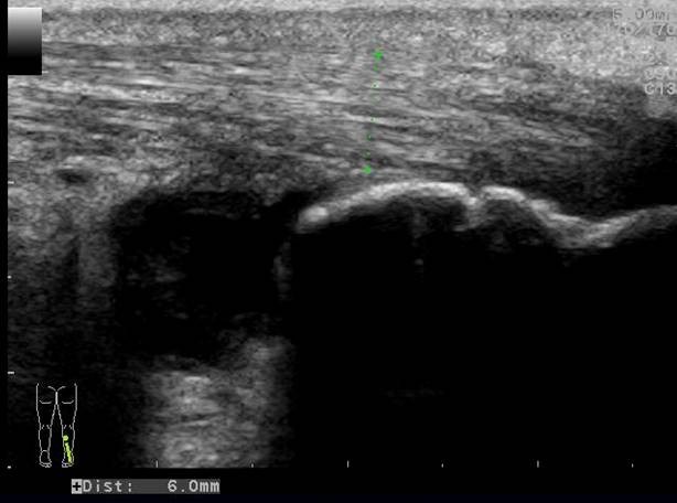 Caption: Longitudinal image of the right distal Achilles tendon | Description: The AP diameter of the achilles tendon is 6 mm.
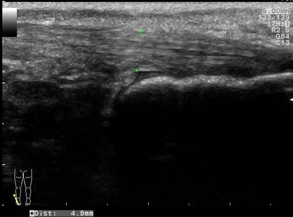 Caption: Longitudinal image of the contralateral (left) distal Achilles tendon | Description: The AP diameter of the normal left achilles tendon is 4,9 mm. There is no fluid filled bursa
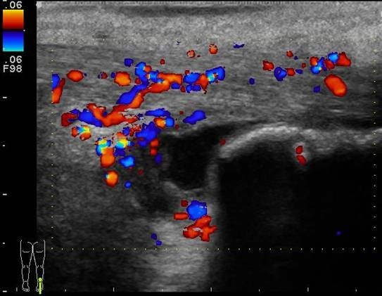 Caption: Longitudinal color doppler image of the right distal Achilles tendon
Description: There is marked hypervascularity in the tendon and bursa wall
Caption: Longitudinal color doppler image of the right distal Achilles tendon
Description: There is marked hypervascularity in the tendon and bursa wall
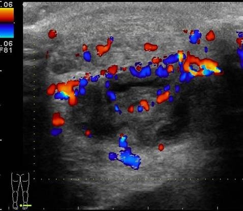 Caption: Transverse color doppler image of the right distal Achilles tendon | Description: There is marked hypervascularity in the tendon and bursa wall
Final Diagnosis
Retrocalcaneal bursitis and Achilles tendon tendinosis
Discussion
There is a thick walled fluid filled bursa between the Achilles tendon and the calcaneal bone. The color Doppler images show hypervascularity of the bursal wall.
A minimal amount of fluid in the retrocalcaneal bursa can often be found.
A retrocalcaneal bursitis is caused by friction of the Achilles tendon over the upper part of the calcaneal bone. It is often an overuse injury found in athletes.
Signs indicating a bursitis are
A rounded contour
A thick wall
Hypervascularity
Increased echogenicity of Kagers fat
A retrocalcaneal bursitis is often found in combination with a tendinosis or tendinopathy of the distal Achilles tendon. Common signs of an Achilles tendinosis or tendinopathy are
Swelling of the tendon
Inhomogenous hypoechoic aspect
Loss of the normal fibrillar pattern
Hypervascularity or neovascularity
In this case there is only slight swelling of the tendon and the normal fibrillar pattern is preserved. There is however a marked hypervascularity.
For more cases of retrocalcaneal bursitis or Achilles tendon pathology visit www.ultrasoundcases.info
Follow Up
Treatment of a retrocalcaneal bursitis is usually conservative with cessation of the sports activities. An ultrasound guided puncture of the bursa and injection of a combination of a corticosteroid and an analgesic can quickly relieve the patient pain
Case ReferencesSchepsis AA, Jones H, Haas AL.Achilles tendon disorders in athletes. Am J Sports Med. 2002 Mar-Apr;30(2):287-305.