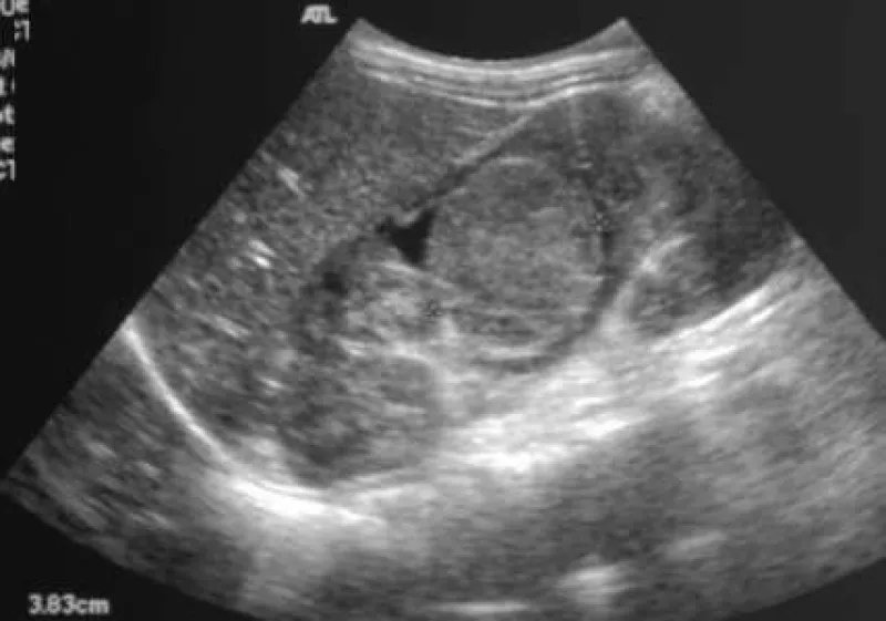
The presence, appearance, and significance of finding cholesterol polyps, adenomyomatosis, within the gallbladder is discussed and reviewed on multiple imaging modalities.
Case stories that stick
The sonographic findings associated with pyloric stenosis are outlined and imaging showing a hypertrophic pyloris are provided in grey scale imaging and with color doppler.
The clinical presentation and imaging results are discussed for a patient whose symptoms of jaundice, intrahepatic and extrahepatic biliary dilitation, and right upper quaderant pain are consistent with a diagnosi...
A structure, referred to as a pseudokidney, is imaged and compared to CT imaging, showing the extent of the disease process of intestinal lymphoma.
Images depicting a micro gallbladder, an ideopathic condition associated with cystic fibrosis, are presented and other possible biliary complications are noted.
Imaging from a man with right lower quaderant pain reveals a bullseye shaped structue with increased blood flow at the area of maximal tenderness, consistent with a finding of acute appendicitis.