
Wilms tumor

First published on SonoWorld
Case Presentation
A previously healthy two and a half year old child presented to primary care physician with upper respiratory symptoms. On examination he was found to have an abdominal mass. An ultrasound and CT scan were performed.
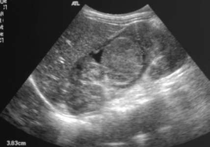 Caption: Sagittal image of the right kidney | Description: A rounded solid appearing mass is seen in the interpolar region of the right kidney. The mass shows no obvious calcifications.
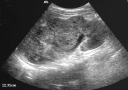 Caption: Sagittal image of the right kidney | Description: The right kidney appears enlarged and lobulated. The complex right renal mass is appreciated again in this image. The mass exhibits a few scattered areas that are hypoechoic.
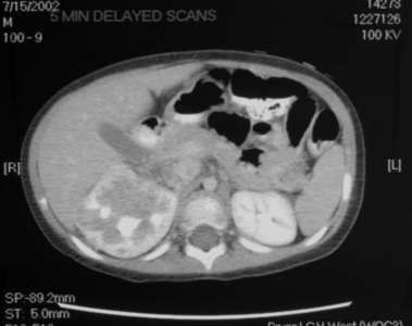 Caption: Contrast enhanced axial CT scan | Description: A relatively non-enhancing right renal mass is seen in this image.
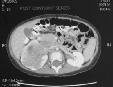 Caption: Contrast enhanced axial CT scan | Description: The right renal mass is again identified in this image. The mass is seen to occupy a large portion of the kidney and invades the right renal hilum; however it does not cross the midline.
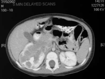 Caption: Contrast enhanced axial CT scan | Description: The right renal mass extends beyond the renal capsule.
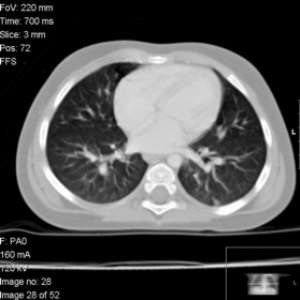 Caption: CT scan of the chest | Description: There is no evidence of pulmonary parenchymal metastases.
Differential Diagnosis
Wilms tumor, duplex kidney, solitary kidney, crossed ectopia, renal contusion/laceration, acute pyelonephritis, acute obstructive nephropathy, nephroblastomatosis, mesoblastic nephroma, multilocular cystic nephroma, ADPKD, renal transplant rejection, medullary sponge kidney, xanthogranulomatous pyelonephritis, acute renal vein thrombosis
Final Diagnosis
Wilms tumor
Discussion
Introduction: Most common primary renal tumor in childhood; peak age 2-3 years; 75% occur before the age of 5
- Arises in nephrogenic blastema
- Epithelial, stromal, blastemal ( undifferentiated components)
- May contain primitive cartilage, osteoid, fat
Histology: Favorable – 88%, Cure rate – 90%
- Anaplastic – 4%: Cellular atypia, more common in older children and African Americans
- Clear cell sarcoma – 6%: “Bone metastasizing renal tumor of childhood,” Mortality – 50%, Bone mets – 40%, may be cystic
- Rhabdoid tumor – 2%, Mortality – 90%, mean age 13 months
Clinical symptoms: Abdominal mass, fever, microscopic hematuria, hypertension
Associations: Hemihypertrophy of any cause; Beckwith-Weidman Syndrome, spontaneous aniridia, increased incidence in crossed renal ectopy and horseshoe kidney
Imaging: Usually homogenous tumor with areas of necrosis, calcification is uncommon (5-10%), may have a cystic component.
Follow Up
An open biopsy was performed which confirmed the diagnosis of Wilms tumor. The patient was started on chemotherapy.