Read this interesting case of a term infant who contracted a Covid-19 infection shortly after birth.
Case stories that stick


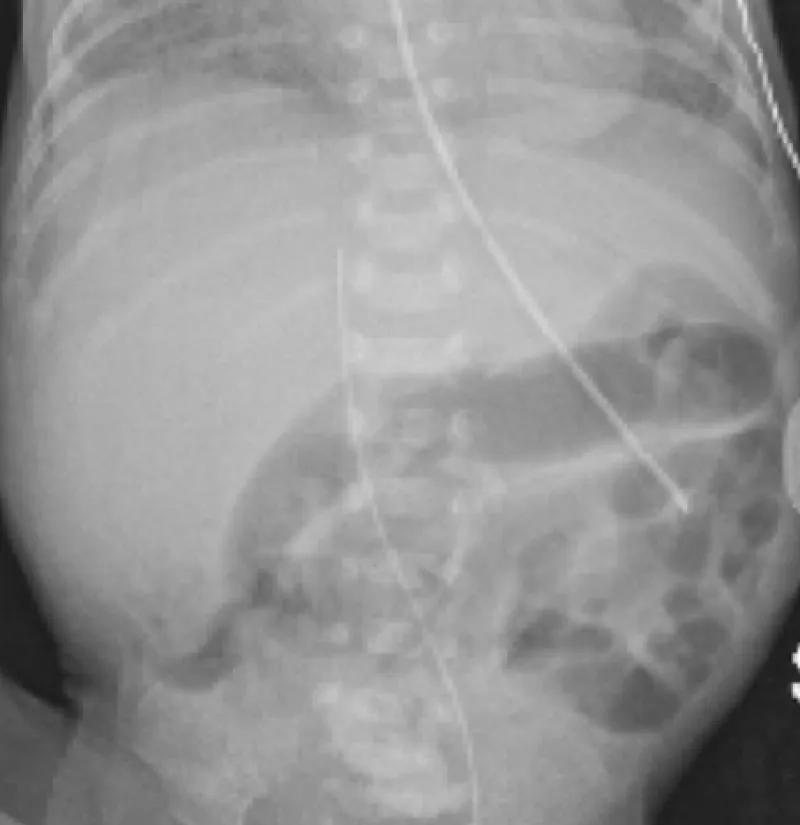
A preterm infant presented with a new onset of emesis and bloody stool – what could be the diagnosis?

External versus internal carotid artery

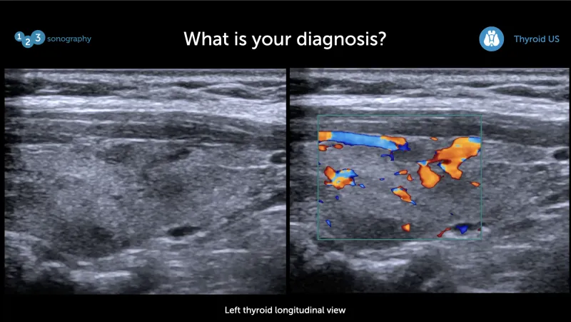
Can you spot the problem on this thyroid ultrasound of a patient with symptoms of hyperthyroidism?
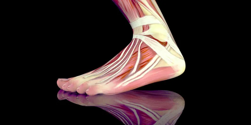
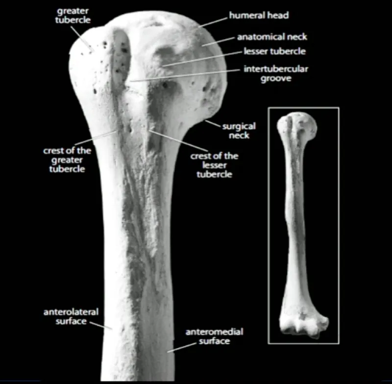
One very important structure to scan within the shoulder protocol is the long head of the biceps. Before scanning you should understand the anatomy of the long head of the biceps. So let´s recap!

MSK Ultrasound is the imaging modality of choice for tendon abnormalities and it is easier than you might think.