Cardiologists are increasingly seeing patients with adult congenital heart disease (ACHD) in their practices. It is now essential for cardiologists, who used to focus on acquired heart diseases, to be knowledgeable ...
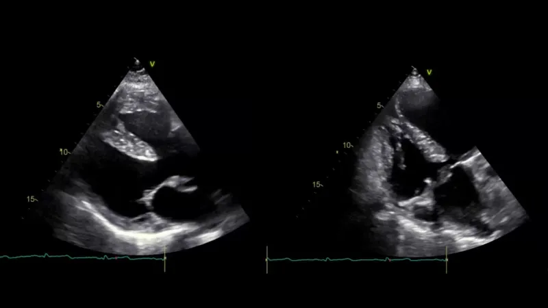
A 70-year-old man presented to the emergency department. Can you spot the diagnosis based on the clinical presentation, laboratory findings, and echocardiographic imaging?
Tue, 21/01/2025
Sophie Wieser, MD
and Prof. Thomas Binder, MD, FESC
Keep in mind: Pleural effusions and consolidations can often be directly or indirectly associated with malignancies—whether lung cancer itself or metastasis from other types of cancer.
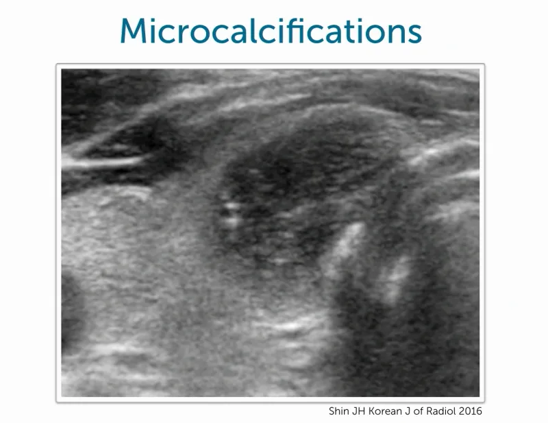
When evaluating thyroid nodules, certain "red flag" features, as categorized by the TIRADS classification, can indicate a heightened risk of malignancy. In this blogpost, we are highlighting prominent indicators tha...