
6. Spectral Doppler

6.1 When should spectral Doppler be applied in carotid ultrasound?
Spectral Doppler is part of every standard carotid scan. In carotid ultrasound we use pulsed wave Doppler. In contrast to continuous Doppler (CW-Doppler), which is mainly used in echocardiography, spectral Doppler allows the measurements of flow velocities within in a sample volume. Since flow velocities are elevated in stenosis, and because these flow velocities increase proportionally with the degree of stenosis spectral Doppler allows us to quantify stenotic lesions.
| Applications of Spectral Doppler |
|---|
| Differentiation ECA from ICA |
| Measure flow velocities |
| To determine if a stenosis is heamodynamically significant |
| Quantification of stenosis |
| Determine the direction of flow |
| See if distal (intracranial) flow obstruction is present |
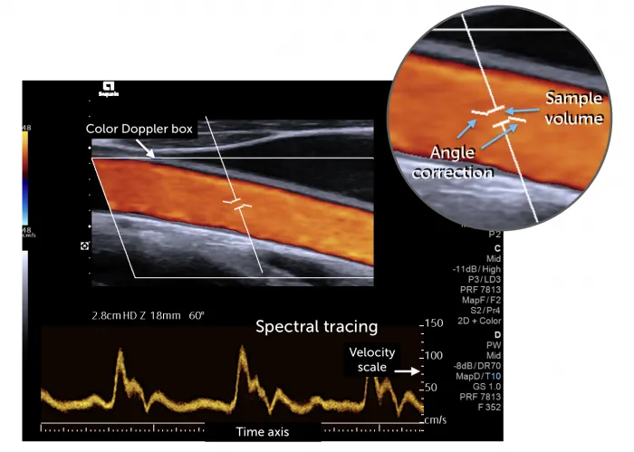
6.2 What is the sample volume in spectral Doppler?
The sample volume is the region where the velocities are measured. This region can be adjusted with the help of the trackball and is depicted as an interrupted line (Doppler line). The width of the sample volume can also be adjusted. The wider the sample volume is the more spectral broadening you will have. An additional line denotes the Doppler angle correction.
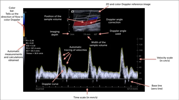
6.3 How should I optimize the spectral Doppler tracing for carotid ultrasound
- Optimize the velocity scale and the base line (shift)
- The curve should be above the baseline (if not invert scale)
- Use a time scale which allows you to observe at least 3 beats at once
- Position your sample volume in the middle of the artery
- The sample volume should be between 15 and 30mm
- Optimize the angle correction (45-60°)
- If you lose the signal - go back to the B mode/color image and try again
- Use automatic curve tracing only if the signal quality is good and tracking is optimal
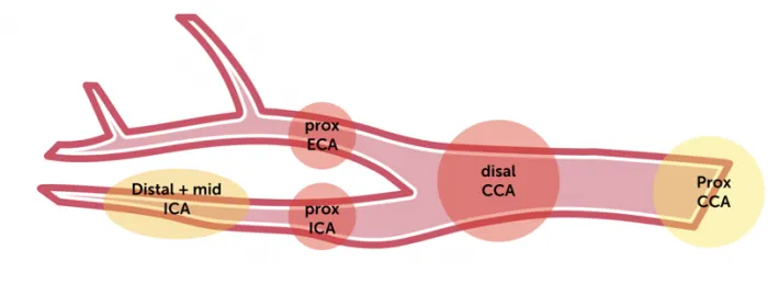
6.4 At which locations should spectral Doppler be performed in carotid ultrasound?
Spectral Doppler measurements should be performed in the common carotid artery, the proximal internal and external carotid artery. It is advisable too also obtain signals from the proximal common carotid artery and the distal + mid carotid artery. It is mandatory to also measure the flow velocity at the site of a suspected stenosis (look for the maximal velocity) and distal to the stenosis.
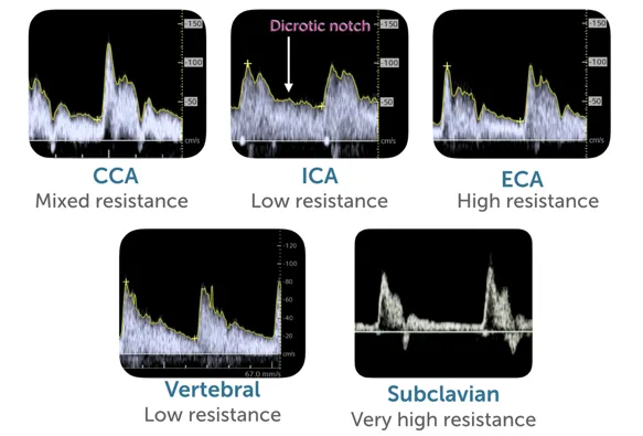
6.5 What are the Doppler characteristics of the arteries of the neck?
Spectral Doppler allows us to identify vessels. Not only is it possible to distinguish arteries from veins but we can also see different flow patterns in different arteries. In general we distinguish high- low and mixed resistance arteries. Arteries that supply the brain are usually low resistance while arteries that supply muscles show a high resistance pattern
| Artery | Type | Characteristic |
|---|---|---|
| Common carotid artery | Mixed resistance | Intermediate systolic / diastolic ratio |
| Internal carotid artery | Low resistance | Lowest systolic to diastolic ratio |
| External carotid artery | High resistance | Highest systolic / diastolic ratio, rapid dip in the curve after systole |
| Vertebral artery | Low resistance | Similar to ICA (low systolic / diastolic ratio) |
| Subclavian artery | Very high resistance | Very little diastolic flow, high systolic / diastolic ration, diastolic flow reversal (triphasic flow) |
6.6 What are the normal flow velocities in the carotid artery?
It is important to measure both end-systolic and diastolic velocities. There is a fairly high variation of the maximal velocities in the arteries of normal individuals. The velocities depend on age, sex, heart rate, stroke volume, resistance and the width of the artery. The following table shows the upper limits of normal that have been proposed in the literature.
Normal values
| Common carotid artery | ||
|---|---|---|
| Peak systolic velocity | < 125cm/s (30-40cm/s) | Large variations in velocity, higher velocities found in paediatric population (up to 180cm/s) |
| End diastolic velocity | 40cm/s? | No data available but usually less than 40cm/s |
| Internal carotid artery | ||
|---|---|---|
| Peak systolic | < 125cm/s | Usually below 100cm/s |
| Enddiastolic | < 40cm/s | Low diastolic velocities are indicative of a distal obstruction |
| Ratio | ||
|---|---|---|
| ICA / CCA ratio | < 2 | Women tend to have slightly higher ratios than men, the ratio increases with age, usually the ratio is between 0.4 - 1.4 |
6.7 Which ratios / calculations are used to quantify the degree of carotid artery stenosis?
At the site of a stenosis (<50%) we see elevated systolic and diastolic velocities. The absolute velocities allow us to quantify the degree of stenosis. In addition we can also compare velocities and calculate ratios. The following ratios are used in carotid artery ultrasound:
| Ratios | |
|---|---|
| ICA/CCA ratio | Diagnose and quantify stenosis |
| S/D ratio | Helps to distinguish high from low resistance vessels, helps to diagnose and quantify stenosis |
| Resistance index | Is calculated as: (peak systolic velocity – end diastolic velocity)/peak systolic velocity. A value of of 1 indicates systolic blood flow but no diastolic blood flow (high resistance vessel) |
| End diastolic ratio ICA/CCA | Helps to quantify the degree of stenosis |