
5. Color Doppler imaging of the carotid arteries


/* .lity-iframe-container iframe { background: #fff !important; } */ .articles__categories nav { display: none !important; } .sonopl-book__aside .articles__categories { background: #f5f5f5; } .sonopl-article th, .sonopl-article td { color: black; text-align: left; } function advagg_mod_1() { // Count how many times this function is called. advagg_mod_1.count = ++advagg_mod_1.count || 1; try { if (advagg_mod_1.count <= 40) { var getClass = document.querySelectorAll(".articles__categories"); var a = document.createElement('a'); var linkText = document.createTextNode("Get Carotid MC Free Lectures"); a.appendChild(linkText); a.title = "newsletter"; a.href = "/overlay/forms/cu_mc/16618"; a.setAttribute("class", "sonopl-button sonopl-button--default"); a.setAttribute("data-lity", ""); a.style = "line-height: 1.2;margin-top: 25px;padding: 15px;width: 100%;"; for(var ix = 0; ix < getClass.length; ix++) { getClass[ix].appendChild(a) } // Set this to 100 so that this function only runs once. advagg_mod_1.count = 100; } } catch(e) { if (advagg_mod_1.count >= 40) { // Throw the exception if this still fails after running 40 times. throw e; } else { // Try again in 250 ms. window.setTimeout(advagg_mod_1, 250); } } } function advagg_mod_1_check() { if (window.jQuery && window.Drupal && window.Drupal.settings) { advagg_mod_1(); } else { window.setTimeout(advagg_mod_1_check, 250); } } advagg_mod_1_check();
5.1 How is color Doppler applied in carotid ultrasound?
Doppler ultrasound is a modality, which allows us to study blood flow in the vessels during carotid ultrasound. It is performed as part of a standard carotid ultrasound exam. Both spectral and color Doppler provide information on blood flow velocity and the direction of flow. Doppler information must always be combined and interpreted together with the B mode image.
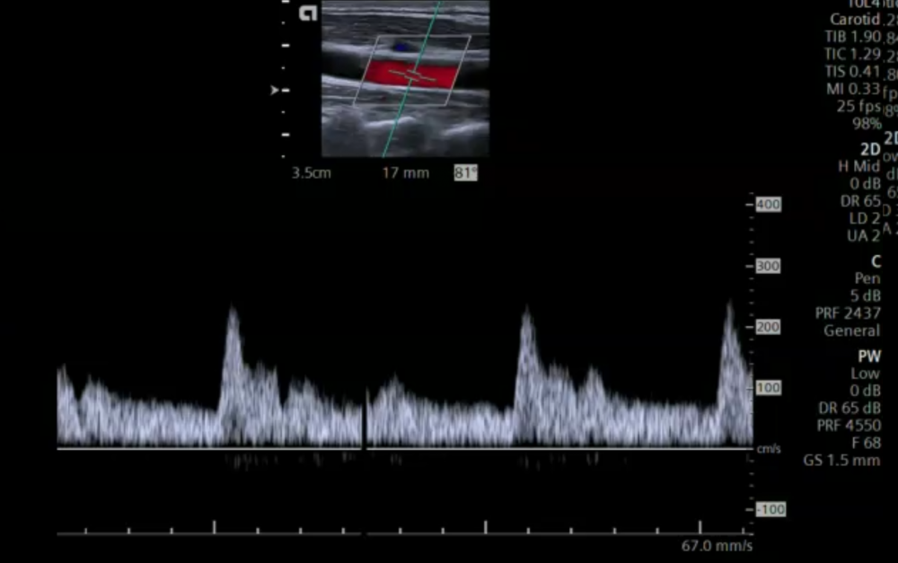 Spectral Doppler (pulsed wave Doppler) tracing in the common carotid artery. The direction of flow can be determined based on whether or not the spectrum is positive (above the zero line) or negative (below the zero line). The x-axis depicts the time. The absolute velocities can be read from the scale on the right (y-axis)
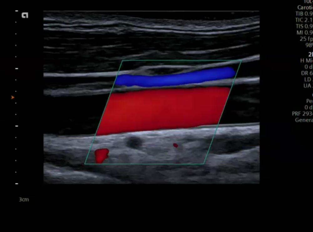
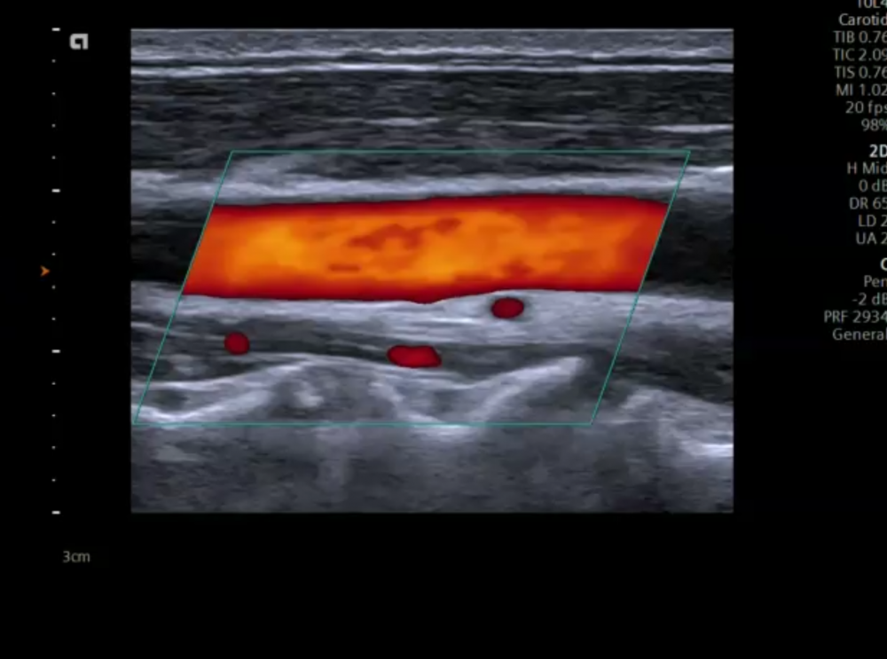 Color –Doppler ultrasound image of the common carotid artery. The Doppler information is provided within a region of interest (ROI) or color box. Depending on the angulations of the color box and the blood flow direction, blood flow is displayed either in red or in blue.
Color Doppler / Duplex provides flow information within a region of interest (ROI), while spectral Doppler provides information within a “sample volume”. Doppler is primarily used in longitudinal views but can also useful in a transverse view.
Color –Doppler ultrasound image of the common carotid artery. The Doppler information is provided within a region of interest (ROI) or color box. Depending on the angulations of the color box and the blood flow direction, blood flow is displayed either in red or in blue.
Color Doppler / Duplex provides flow information within a region of interest (ROI), while spectral Doppler provides information within a “sample volume”. Doppler is primarily used in longitudinal views but can also useful in a transverse view.
5.2 How is Doppler used in a transverse view?
The transverse view does not allow us to determine the direction of flow. Therefore PW Doppler is rarely used in a transverse view (we can not correct the Doppler angle). However, there are several ways how color Doppler can be applied:
Color Doppler in a transverse view Can be used to spot turbulences To see if the entire vessels fills (excluding or detecting soft plaque) Can help to discern arteries from veins Can help to discern internal from external carotid artery Can help to follow vessels during a sweep Can help to detect tortuous vessels Use a rectangular color box Angulate transducer caudally or cranially to get a suitable Doppler angle (avoid a mixed red and blue signal) Adjust PRF Also try Power Doppler
5.3 What is Power Doppler and how is it used in carotid ultrasound?
Power Doppler is an ultrasound modality, which uses only the amplitude of the Doppler signal to display flow. The Doppler intensity is displayed in colors from red (low intensity) to yellow (high intensity). Power Doppler is angle independent and is not affected by aliasing. In contrast to color Doppler Power Doppler does not display the direction of flow. In general it is more sensitive for lower flow velocities and flow in small vessels. Power Doppler can also be used to examine vessels in superficial organs such as the thyroid or testicles and to visualize vessels in tumors.
Power Doppler Power Doppler uses the amplitude of the Doppler signal to display flow More sensitive for the detection of blood flow than color Doppler We loose the information of blood flow direction There is no aliasing It is less angle dependant Allows visualization of small vessels It can be used to visualize the inner contour of the vessels and to detect or exclude areas of narrowing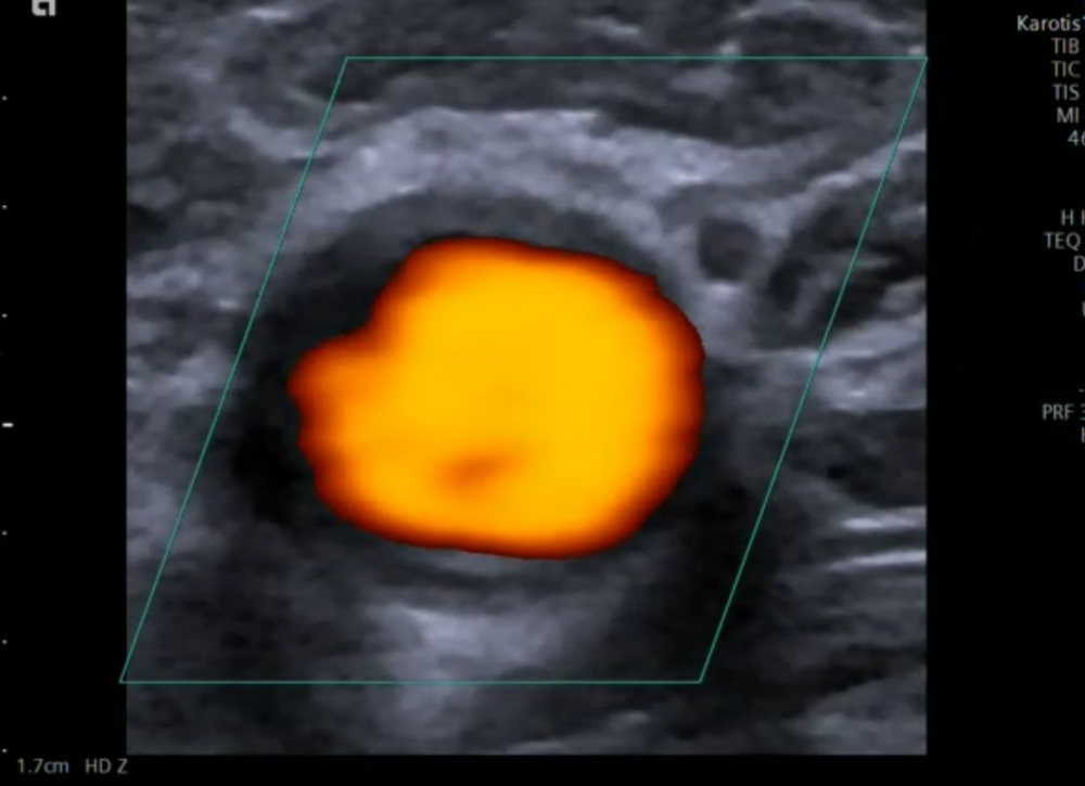 Transverse view of the common carotid artery a small posterior soft plaque can be seen. Power Doppler us used to delineate the wall. No color is seen in the region of the plaque.
5.4 How is Doppler used in a longitudinal view?
Color Doppler is used to demonstrate the direction of flow and to depict areas of higher flow velocities. Higher flow velocities are characterized by aliasing. Color Doppler (together with power Doppler) is also helpful to depict plaques with low echogenicity (soft plaque), which might be missed with B mode imaging alone. Spectral Doppler allows characterization of the vessel (i.e. vein vs. artery, internal vs. external carotid artery) and measure flow velocities (systolic and diastolic), which are then also used to calculate indices. Flow velocities allow us to quantify the degree of a stenotic lesion and to determine if such a lesion is relevant or not.
5.5 What is the color Doppler Box and how should it be adjusted during carotid ultrasound?
The color box is the region of interest in which color flow information is displayed. The size, position and angle of the color box can be adjusted.
Color Box The color box is the region of interest where color flow will be depicted The scale will tell you what the PRF (aliasing limit) and the direction of flow is (you can invert the color scale) Tilt the transducer (caudal and cranial) to optimize your angle of insonation (Try not to be parallel to the vessel with your imaging orientation - as in A) The color box angle can be adjusted so that you are at an optimal angle to blood flow The diagonal (long axis) of the box should be as parallel as possible to the vessel wall No angle correction is required if flow is directly towards or away from the transducer (parallel to flow) If the color box is optimized your color signal will be much betterThe orientation of the color box should follow the direction of flow. If the Doppler interrogation is perpendicular Doppler does not allow us to determine the direction of flow. This means you will get a mixed red / blue signal. Try to use a flow orientation where the Doppler angle is as parallel to blood flow as possible. This can be achieved by changing the angle of insonation and by adjusting the color box. The long axis of the box should approximate the direction of flow
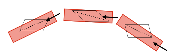 Three different vessel / flow orientations. The color box should be adjusted accordingly to achieve best results. – note that the longest diagonal of the box approximates the direction of flow.
 Color Doppler displays both the flow in the common carotid artery (red) and the jugular vein (blue) note that the color box angle is adjusted so that its “long diagonal” approximates the direction of flow
5.6 How should color Doppler be adjusted?
It is important not only to adjust the size and angle of the color box, but also to adjust the color gain, set the aliasing velocity (PRF) and to chose an appropriate color map. Here are some important tips:
Color Doppler tips First optimize your PRF (CCA). Choose a velocity which is just below the Nyquist limit (aliasing) Primarily use conventional Doppler, add Power Doppler in specific situation Always use the same maps (chose one that fits you), Optimize you color Box Optimize colors gain (avoid high gain settings, which cause blooming artifacts) Observe the direction of flow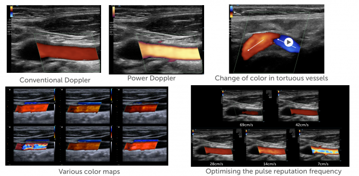 Top left: Difference between conventional color Doppler and power Doppler. Top right: note how the color changes from blue to red once the flow direction changes.
Bottom left. Different color maps. Bottom right: Effect to the pulse repetition frequency (PRF) on the color Doppler display. A low PRF will cause aliasing
Top left: Difference between conventional color Doppler and power Doppler. Top right: note how the color changes from blue to red once the flow direction changes.
Bottom left. Different color maps. Bottom right: Effect to the pulse repetition frequency (PRF) on the color Doppler display. A low PRF will cause aliasing
5.7 What is recirculation in carotid ultrasound?
Recirculation Recirculation which is also called „flow separation“ is a specific flow pattern found in the bulb Lower peak systolic velocity flow Circulating and retrograde flow The magnitude varies from patient to another It has been hypothesized that the flow dynamics of the bulb play a role in the likelihood for developing plane and stenosis The presence of recirculation can help to identify the region of the bulb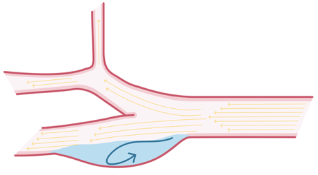 Recirculation is a typical flow pattern found at the bulb. Recirculation is caused by widening of the artery at the bulb. Here you will see retrograde/circulating of flow
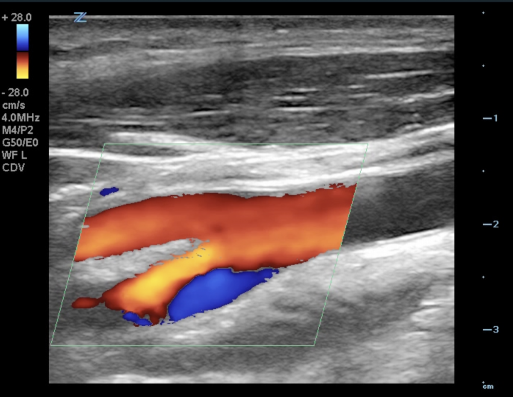 Carotid bifurcation of a healthy individual. The internal carotid artery is seen dorsal, the external carotid artery is located ventral. Note the area of recirculation (blue color Doppler flow signal) in the carotid bulb (internal carotid artery)
.pagination .pager .pager__item { font-size: 12px; } .pagination .pager .pager__item--current { padding: initial; } .pagination { margin: 0; margin: 0 auto; margin-top: 0 !important; } .pager { margin-top: 0 !important; padding: 0; display: flex; } .pagination .pager { padding: 0; } .pagination .pager a { padding: 8px 12px; } .pager__item a { box-shadow: none !important; } .aut-nav-carotid-p { text-align: center !important; margin-bottom: 6px !important; } function advagg_mod_2() { // Count how many times this function is called. advagg_mod_2.count = ++advagg_mod_2.count || 1; try { if (advagg_mod_2.count <= 40) { var autNaviCarotid1 = ('\ \
- \
- \
Chapters\
\
- \ 1\ \
- \ 2\ \
- \ 3\ \
- \ 4\ \
- \ 5\ \
- \ 6\ \
- \ 7\ \
- \ 8\ \
- \ 9\ \
- \ 10\ \
- \ 11\ \
- \ 12\ \
- \ 13\ \
- \ 14\ \
- \ 15\ \
- \ last »\ \ \ \ '); var autNaviCarotid2 = ('\
Carotid Ultrasound Webbook & Wiki\ \
- \
- \
‹ previous\
\
- \ BACK TO OVERVIEW\ \
- \ next ›\ \ \ \ '); (function($) { $(document).ready(function(){ $(".sonopl-article.sonopl-book__content.sonopl-content-main") .prepend(autNaviCarotid1) .prepend(autNaviCarotid2); $(".aut-nav-carotid") .prepend(autNaviCarotid1) .prepend(autNaviCarotid2); }); }(jQuery)); // Set this to 100 so that this function only runs once. advagg_mod_2.count = 100; } } catch(e) { if (advagg_mod_2.count >= 40) { // Throw the exception if this still fails after running 40 times. throw e; } else { // Try again in 250 ms. window.setTimeout(advagg_mod_2, 250); } } } function advagg_mod_2_check() { if (window.jQuery && window.Drupal && window.Drupal.settings) { advagg_mod_2(); } else { window.setTimeout(advagg_mod_2_check, 250); } } advagg_mod_2_check();

If you like the way we teach, please leave a message!
- \ BACK TO OVERVIEW\ \
- \ 1\ \