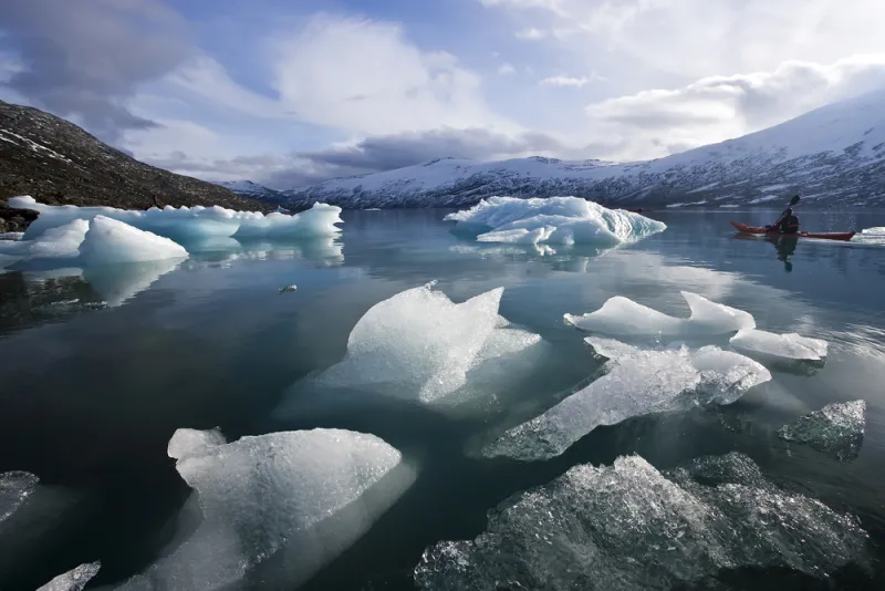
Frozen Stream Nature provides several opportunities to understand echocardiographic findings. Thu, 17/12/2020 Univ. Prof. Dr. Thomas Binder
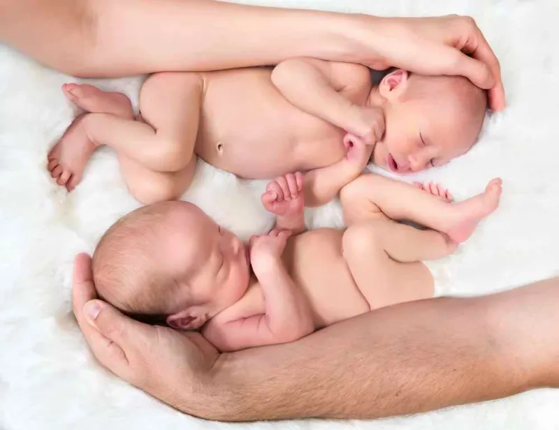
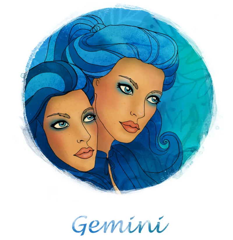
Gemini Part 2 Remember one of the last cases in which I wrote about heart failure and mitral regurgitation? Well, here is the sequel... Wed, 16/12/2020 Univ. Prof. Dr. Thomas Binder
Gemini Part I As doctors we are usually responsible for the life of ONE patient. However, this is not always the case... Wed, 16/12/2020 Univ. Prof. Dr. Thomas Binder
Echo Wiggles Ultrasound case: Mr. Kaminski had never been seriously ill. He had always taken good care of his health. But then ... Wed, 16/12/2020 Univ. Prof. Dr. Thomas Binder
Echo The Paradox Adhering to rules gives us a certain sense of confidence. The same is true for echocardiography. Wed, 16/12/2020 Univ. Prof. Dr. Thomas Binder
Echo Guinness Book of Echo I It is part of human nature to be fascinated by extremes, by the unusual or the bizarre. Why should it be different in echocardiography? Wed, 16/12/2020 Univ. Prof. Dr. Thomas Binder