
16.7 Right heart involvement in systemic diseases
The right ventricle may be involved in many systemic diseases. The following section will provide a short overview of those pathologies that may affect the right ventricle and list specific characteristics and features that may be found. A more detailed description will be provided in the chapters dealing with the specific diseases.
16.7.1 Myocarditis
Myocarditis does not affect the left ventricle alone; it may involve the right ventricle as well. Isolated right ventricular myocarditis has also been described. The principal findings are impaired right ventricular function and pericardial effusion.
16.7.2 Right ventricular dysfunction in cardiomyopathy
Right ventricular function may be impaired not only due to left heart failure, but also because the right ventricular myocardium is involved in the initial disease process (biventricular cardiomyopathy). Reduced right ventricular function in cardiomyopahy is associated with a poor prognosis.
16.7.3 Right ventricular involvement in eosinophilic syndromes
The hallmark of this disease is the presence of endomyocardial fibrosis and eosinophilic infiltrates in the apex. Infiltrates may also be present in the right ventricular apex. Additionally one may observe right ventricular dysfunction and increased echogenicity of the endocardium of the right ventricle.
Video Platform
Video Management
Video Solutions
Video Player Endomyocardial fibrosis with eosinophilic infiltrates in both ventricles (apex)
16.7.4 Right ventricular involvement in infiltrative heart disease
Sarcoid heart disease and amyloidosis may affect both ventricles. Infiltrates may be found in the right ventricle as well. In amyloidosis, right heart involvement is found in early stages of the disease. Right heart failure is the predominant clinical symptom in patients with amyloid heart disease.
16.7.5 Right ventricular involvement in hypertrophic cardiomyopathy
Right ventricular mass is frequently increased in hypertrophic cardiomyopathy. Approximately one third of patients have severe hypertrophy with a free wall thickness (measured at the free lateral wall of the right ventricle) exceeding 15 mm. A subset of patients may even develop outflow tract obstruction in the right ventricle.
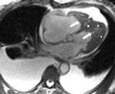
16.7.6 Thorax deformities
The right ventricle is positioned anteriorly and may therefore be affected by abnormalities of the chest, such as pectus excavatum (funnel chest), in which the sternum and the ribs are sunken. The right ventricle is compressed and slim. The heart may be displaced to the left.
Patients with abnormalities of the chest are difficult to image. Use atypical views.Hemodynamic impairment of the heart is observed only in severe forms. In this condition one may find restrictive filling patterns and flow acceleration at the level of the tricuspid valve annulus.
Video Platform
Video Management
Video Solutions
Video Player Compressed right ventricle in a patient with a funnel chest
16.8 General remarks
Assessment of the right ventricle is a challenging subject. Even experienced investigators may find it difficult to correctly assess the size and function of the right ventricle. However, to "sense" abnormalities of the right ventricle one must look for indirect signs, such as the presence of tricuspid regurgitation, the width of the inferior vena cava, and the motion of the interventricular septum. New modalities like speckle tracking and three-dimensional echocardiography will expand our knowledge of the physiology of the right ventricle and help to diagnose its pathologies. Ultimately, all findings must be interpreted in the respective clinical context.