
16.2.3 General approach to pulmonary hypertension
Echocardiography is the primary screening modality to identify pulmonary hypertension. The diagnosis is not always simple. Subtle forms require an integrative approach, especially when tricuspid regurgitation is absent. The patients may present with atypical symptoms. Other modalities may provide important clues concerning the presence of pulmonary hypertension (such as increased NT Pro-BNP levels, right ventricular strain pattern on ECG). Patients who are at high risk for pulmonary hypertension (see list) should be given special attention.
Drugs that cause or may cause pulmonary hypertension Aminorex Fenfluramine Dexfenfluramine Toxic rapeseed oil Benfuorex Amphetamines L-tryptophan Metamphetamines Cocaine Phenylpropanolamine Chemotherapeutic agents Selective serotonin re-uptake inhibitors Pergolie Predisposing conditions Connective tissue disease (especially systemic sclerosis) Deep vein thrombosis, recurrent pulmonary embolism Congenital heart disease Familial history of pulmonary arterial hypertension Portal hypertension HIV infection Osler Weber Rendu syndrome Thalassemia Several treatment options are available for patients with pulmonary hypertension. These include treatment of the underlying cause (such as left heart disease), surgical thrombectomy (for CTEPH), and specific drugs to reduce pulmonary artery pressure (e.g. endothelin receptor antagonists, phosphodiesterase 5 Inhibitors, prostanoids).Patients should be followed on a regular basis. Always exclude left heart disease as the cause of pulmonary hypertension (post-capillary hypertension). Many elderly patients tend to have some degree of pulmonary hypertension. All other patients require invasive hemodynamic assessment. By definition, the diagnosis can be confirmed only by direct measurement of pulmonary pressure. Echocardiography provides significant information in respect of the severity and effects of pulmonary hypertension. Specifically, echocardiography should be used to assess the prognosis and monitor the effects of treatment.
Pulmonary hypertension must be diagnosed early because advanced forms are less responsive to treatment.16.3 Acute pulmonary embolism
The reaction of the right ventricle to acute pressure overload, as in acute pulmonary embolism, differs from that in chronic pressure overload. The right ventricle does not have time to adapt and cope with the sudden high afterload. Thus, it will not be able to generate high pressures. This is the reason why pulmonary embolism is not associated with significant pulmonary hypertension. Systolic pulmonary pressure rarely exceeds 45-60 mmHg. In fact, the typical reaction of the right ventricle is that of right heart failure (dilated right ventricle with reduced right ventricular function). Very high pulmonary artery pressures are registered only when the patient had previous pulmonary embolism that enabled the right ventricle to adapt to the condition.
Echocardiography is not very sensitive for the detection of minor to moderate pulmonary embolism.Typical echocardiographic findings of pulmonary embolism are usually present only when the patient has at least some degree of hemodynamic impairment.
The most widely propagated diagnostic criterion for the diagnosis of pulmonary embolism is the McConell sign: apical segments of the RV show normal or even hyperdynamic motion while the free lateral wall is hypokinetic. While such a pattern is highly specific for pulmonary embolism, it is not very sensitive. Besides, it may also exist in the presence of right heart infarction.
Video Platform
Video Management
Video Solutions
Video Player Echocardiogram of a patient with severe acute pulmonary embolism. The right ventricle is dilated and the McConnell sign is present.
Another sign is the so called 60/60 sign: pulmonary pressure is below 60 mmHg, and pulmonary valve acceleration time (PVAT) is lower than 60 msec.
When you suspect a pulmonary embolism, actively look for right heart thrombi.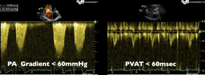
Video Platform
Video Management
Video Solutions
Video Player Patient with acute pulmonary embolism and a large thrombus (following deep vein thrombosis) in the right atrium Always interpret echocardiographic findings in a clinical context when looking for pulmonary embolism.
Echocardiography clearly has certain limitations as regards the diagnosis of pulmonary embolism. The true value of echocardiography lies in the assessment of the hemodynamic effects of pulmonary embolism (i.e. whether the right ventricle is able to cope with the acute pressure load). Very large ventricles, severely reduced right ventricular function, severe tricuspid regurgitation, low right ventricular stroke volume, and pericardial effusion are indicative of more severe pulmonary embolism, and are associated with a poor prognosis. These patients might be candidates for thrombolysis.