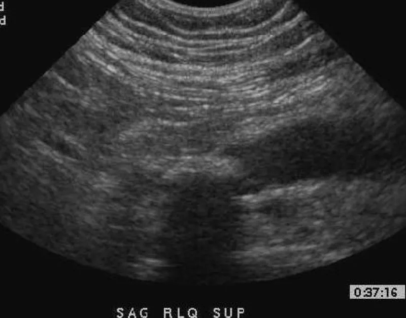
Imaging of the right lower quaderant is provided showing a non-compressable hypoechoic structure with hyperemia and calcification and the diagnosis of acute appendicitis is discussed as well as the process of diag...
Along with a discussion of other possible locations and presentations of metastatic lymph adenopathy, images depicting abdominal metastatic adenopathy and associated ascites are presented in the setting of diffuse...
Imaging from a woman with acute right upper quadrant pain is shown, illustrating a thickened gallbladder wall, cholelithiasis, hyperemia, and pericholecystic fluid, constant with acute cholecystitis.
A grey scale and doppler iterrogation of a TIPS shunt shows a full obstruction in the setting of a patient with hepatic cirrhosis, portal hypertension, and increasing abdominal girth due to ascites.