Metastatic abdominal adenopathy
First published on SonoWorld
Case Presentation
A young man presents with diffuse pain in the abdomen. He gives a history of cutaneous melanoma excised from the right groin. An abdominal ultrasound was performed.
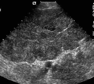 Caption: Transverse image below the level of the umbilicus. | Description: A large, lobulated heterogeneous soft tissue mass is seen in the pelvis.
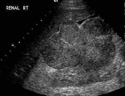 Caption: Sagittal view of the right hypochondrium. | Description: A lobulated, heterogeneous mass is seen in the subhepatic space, abutting the right kidney. The mass also impinges on the right lobe of the liver. A small amount of ascites is seen in this image.
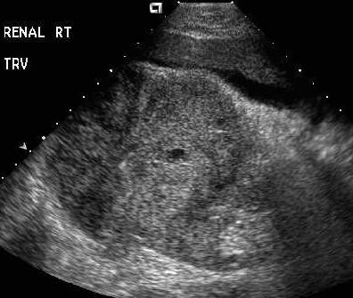 Caption: Transverse view of the right kidney. | Description: No clear fat plane is identified between the mass and the right kidney, suggesting the possibility of the mass infiltrating the kidney. There is no hydronephrosis. The ascites is noted again.
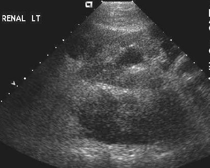 Caption: Transverse view of the left kidney. | Description: The left kidney demonstrates mild hydronephrosis. Multiple hypoechoic masses are noted adjacent to the left kidney [in this image two masses are seen, one anteriorly and one large mass posteriorly].
Differential Diagnosis
1. Metastatic nodes, 2. Infectious adenopathy, 3. Lymphoma [less likely, as lymphoma typically produces hypoechoic masses]
In a patient with known history of melanoma, findings of enlarged nodes, trace ascites and mild left hydronephrosis more likely represent a metastatic process involving the abdomen. The presence of probable invasion of the right kidney by the nodal mass further strengthens this impression.
Final Diagnosis
Multiple, metastatic enlarged nodes throughout the abdomen with trace ascites and mild left hydronephrosis. An MRI scan confirmed the infiltration of the right kidney by the nodal mass.
Discussion
According to Leiter et al, metastases of cutaneous melanoma develop via three main metastatic pathways and occur as satellite or in-transit metastasis, as regional lymph node metastasis or as distant metastasis at the time of primary recurrence. Many metastatic melanomas are silent i.e. they cause no symptoms. Organ failure due to the metastatic process is usually the cause of death in these patients. In its distant spread, melanoma can involve almost any organ; however, the organs commonly involved besides lymph nodes are lungs, liver, brain, kidneys and adrenals.
Metastatic lymph nodes can be detected by ultrasound, CT and MRI. CT is most often the modality used to survey for metastatic disease in patients with a primary cancer. However, because of the specific signal characteristics of melanoma, MRI is frequently used as well.
Ultrasound appearance –
- In the early stages, the metastatic lymph nodes may be identified as discrete, enlarged structures.
- The nodes have variable echogenecity, [a point to be remembered is that these nodes are typically not as hypoechoic as those seen in lymphoma].
- As the nodes enlarge further, they can form conglomerate nodal masses exhibiting a heterogeneous echo pattern.
- Infiltration of the adjacent organs by these nodal masses may also be noted.
- Ascites may be present.
The formation of complex nodal masses in the abdomen points to an advanced stage of the disease. An infectious process such as tuberculosis may have a similar appearance on ultrasound and differentiating it from a metastatic process may be difficult whereas the nodes in lymphoma tend to have a very hypoechoic, almost cystic appearance.
Role of abdominal ultrasound in a case of melanoma–
- Identify the metastatic process and assess its extent. However, small liver metastases may not be apparent on ultrasound, and CT or MRI are more sensitive.
- Provide guidance for fine needle aspiration.
- Monitor results of therapy by documenting the increase or decrease in nodal size. CT or MRI may be performed for the same purpose.
Follow Up
This patient had a biopsy which established the diangosis of metastatic melanoma involving the abdominal nodes.
Case References
1.Leiter U, Meier F, et al. The natural course of cutaneous melanoma. J Surg Oncol. 2004 Jul 1; 86(4):172-8.
2.Rumack, et al. the retroperitoneum and great vessels. Diagnostic ultrasound, second edition, Chapter 12: 457-460.

