
4.2 Physiology of diastole
4.2.1 Timing and phases of diastole
Filling of the ventricle occurs during diastole, which is defined as the time interval from closure of the aortic valve to termination of mitral inflow. On the ECG diastole is crudely defined by the time interval between the end of the T-wave and the QRS complex.
Echocardiography is more accurate than ECG in determining the duration of diastole, and also permits the investigator to distinguish between the different phases of diastole. Both PW Doppler of mitral inflow and LVOT flow are used to measure diastolic time intervals. Tissue Doppler at the mitral annulus is also used to assess the various phases of diastole. We define the following phases:
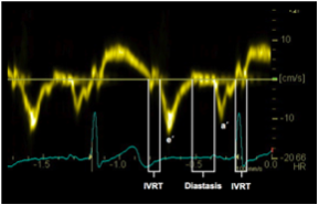
IVRT - Isovolumetric relaxation time - End of LVOT/AV flow to the beginning of the E-wave
Rapid early filling - Duration of the E-wave
Diastasis: Time between the E- and the A-wave
Late atrial filling (atrial contraction) - Duration of the A-wave.
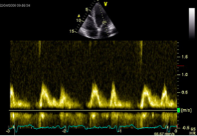
The early phase of filling is caused by the pressure gradient between the atrium and the ventricle. The left atrium is filled while the left ventricle is in a state of relaxation. The pressure gradient is rather small, but still sufficient to open the mitral valve and permit filling. A second important factor of early filling is diastolic recoil of the ventricle. Especially in young and healthy individuals this recoil causes suction, which drags blood from the atria into the ventricle. Filling of the ventricle is also caused by the motion of the heart itself. During diastole the ventricle expands and engulfs the volume of the left atrium. Early filling correlates with the E-wave of transmitral flow. Basically, the size and velocity-time integral of the E-wave reflect the quantity of blood that enters the ventricle during early filling.
Filling of the ventricle causes pressure in the left ventricle to rise. When equilibration of pressure between the atrium and the ventricle occurs, transmitral flow ends and the mitral valve partially closes. This phase, during which little or no filling occurs, is known as diastasis and correlates with the interval between the E- and the A-wave of the transmitral Doppler signal.
The third phase of filling is caused by atrial contraction and is therefore only present in sinus rhythm. Atrial contraction correlates with the A-wave of the mitral inflow signal. The position of the A-wave relative to the QRS complex (the beginning of systole) and its relationship with the E-wave depend on heart rate and the length of the PQ interval. When the AV delay is long (first degree AV block) the A-wave will be closer to the E-wave and farther away from the onset of systole. When it is very short, atrial contraction may coincide with ventricular contraction. In this case, filling will end early.
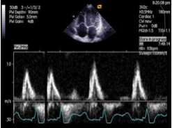
The length of diastole is also influenced by heart rate. An increase in heart rate shortens diastole more than systole. At a higher heart rate diastasis will eventually vanish. The E- and the A-wave will overlap. When heart rate increases even further you will observe complete fusion of the E- and the A-wave.
