
3.4.3 Left atrial size
Left atrial enlargement is either the result of elevated atrial pressure or an increase in flow. However, the degree of left atrial enlargement depends on the compliance of the left atrial wall. This is why you might see patients with fairly high atrial pressures but only modest enlargement of the left atrium, or vice versa. Another important factor that contributes to atrial enlargement is atrial fibrillation. Atrial fibrillation leads to "scarring" of the atrial wall and ultimately to (further) dilatation of the left atrium.
Disproportionate enlargement of the left atrium (in relation to the left ventricle) in athletes may be a clue to the presence of myocardial disease
Video Platform Video Management Video Solutions Video Player Normal LA Video Platform Video Management Video Solutions Video Player Dilated LAIn many cases it will be quite obvious from the 2D image that the atrium is enlarged. If left atrial pressure is also high you will see the interatrial septum bulging to the right and expansion of the left atrial appendage. The atrium may become quite huge, especially in mitral stenosis and restrictive cardiomyopathy. The largest atrium I encountered had a volume of more than 800 ml.
Visual assessment of size provides a first rough estimate. It is essential to perform measurements. Previously, the MMode from a parasternal long axis was used to quantify the left atrium. This diameter represents the anterior posterior dimension of the left atrium. A diameter of < 40 mm and a ratio left atrium/aortic root of < 1.3 are considered normal. The major problem of the MMode is that perpendicular orientation to the left atrium may not be possible. Therefore, 2-D measurements have now replaced the MMode. Measurements should be performed in apical views (four- and two-chamber view) during end-systole.
Avoid foreshortening of the left atrium.
Both the length and the width of the left atrium can be determined here. However, make sure you perform the measurements more or less parallel to the interatrial septum from the plane of the mitral annulus to the roof of the atrium, and do not include the right upper pulmonary vein.
2D measurements are very simple to obtain and fairly reliable. Still, the preferred method to quantify left atrial size is now volumetric methods because they also account for variations in the shape of the left atrium. There are two different principles with which volumes can be obtained: the area-length method and the method of discs (Simpson method). For both methods, tracing of the atrial cavity is required. This is done on a four chamber view at end systole, shortly before the mitral valve opens. For the biplane approach, which is even more exact, also use a 2-chamber view.
Include a septal aneurysm but not the left atrial appendage or the pulmonary veins in your tracing of the left atrium.
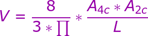
The area length method is based on a geometric assumption and employs the following formula:
The Simpson method uses the rule of discs, which has already been described for the left ventricle (see: 3.2.3.2 2 The Simpson method to determine ejection fraction).
Most measurement programs automatically derive both volume computations (in ml). The area length method is probably a little less exact than the Simpson method, but both can be used because they yield very similar results. All of the measurements can then be indexed to the body surface area.
Inclusion of pulmonary veins Inclusion of tenting area Alignment / foreshortening Lateral resolution not at end-systole PitfallsLeft atrial volume is a strong predictor of events. There is evidence that patients with left atrial volumes above 34 ml per m2 are at high risk for ischemic stroke, heart failure, atrial fibrillation, and death.
Volume calculations can also be performed with 3D technologies. Although this approach is mainly used in research and has not reached clinical practice yet, it might be the method of the future.

 Normal (mm)
30—40
27—38
Mild (mm)
41—46
39—42
Moderate (mm)
47—52
43—46
Severe (mm)
≥ 52
≥ 47
MMode Measurements of LA — Reference Values
Normal (mm)
≤ 50
Mild (mm)
51—60
Moderate (mm)
61—70
Severe (mm)
> 70
LA Length — Reference Values
Normal (cm2)
≤ 20
Mild (cm2)
20—30
Moderate (cm2)
30—40
Severe (cm2)
> 40
LA Area
Normal (mL)
18- 58
22- 52
Mild (mL)
59- 68
53- 62
Moderate (mL)
69- 78
63- 72
Severe (mL)
≥ 79
≥ 73
LA Volume
Normal (mL/m2)
22±6
Mild (mL/m2)
28-33
Moderate (mL/m2)
34-40
Severe (mL/m2)
> 40
Indexed LA Volume
Normal (mm)
30—40
27—38
Mild (mm)
41—46
39—42
Moderate (mm)
47—52
43—46
Severe (mm)
≥ 52
≥ 47
MMode Measurements of LA — Reference Values
Normal (mm)
≤ 50
Mild (mm)
51—60
Moderate (mm)
61—70
Severe (mm)
> 70
LA Length — Reference Values
Normal (cm2)
≤ 20
Mild (cm2)
20—30
Moderate (cm2)
30—40
Severe (cm2)
> 40
LA Area
Normal (mL)
18- 58
22- 52
Mild (mL)
59- 68
53- 62
Moderate (mL)
69- 78
63- 72
Severe (mL)
≥ 79
≥ 73
LA Volume
Normal (mL/m2)
22±6
Mild (mL/m2)
28-33
Moderate (mL/m2)
34-40
Severe (mL/m2)
> 40
Indexed LA Volume