3.4.2 Anatomy and function of the left atrium
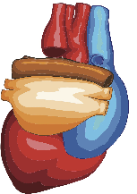
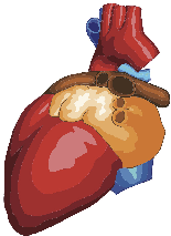
The left atrium consists of three parts: the vestibule, the left atrial appendage, and the pulmonary-vein component. The left atrium is more or less oval-shaped. Its extension is greater in the superior-inferior aspect than in the antero-posterior or medial-lateral aspect. The left atrium is largest at end-systole, shortly before the mitral valve opens. The entrance to left atrial appendage lies in close proximity to the left upper pulmonary vein, from which it is separated by a rather prominent ridge. The left atrial appendage has a finger-like appearance and "rests" on the anterolateral portion of the left ventricle. It is the most common site of thrombi in the left atrium and sensitive to any rise in left atrial pressure. Large quantities of natriuretic peptides are stored and secreted at this site. The interatrial septum divides the left and the right atrium. The oval-shaped fossa ovalis is defined by the region in which the primary septum is not overlapped by the secondary septum. This region is thinner than the remaining portions of the interatrial septum. Usually four distinct pulmonary veins enter the left atrium (left and right, upper and lower pulmonary veins). However, one may well find anatomical variations such as a common trunk with two pulmonary veins entering the left atrium simultaneously, or the presence of additional pulmonary veins.


The wall of the left atrium is rather thin (some parts are only 1.5 to 2 mm thick) and smooth. An exception is the left atrial appendage, which is rough with pectinate muscles.
If you place a flashlight behind the atrial wall you will actually see the light through the left atrial wall. Video Platform Video Management Video Solutions Video Player TEE study displaying the left atrial appendage