


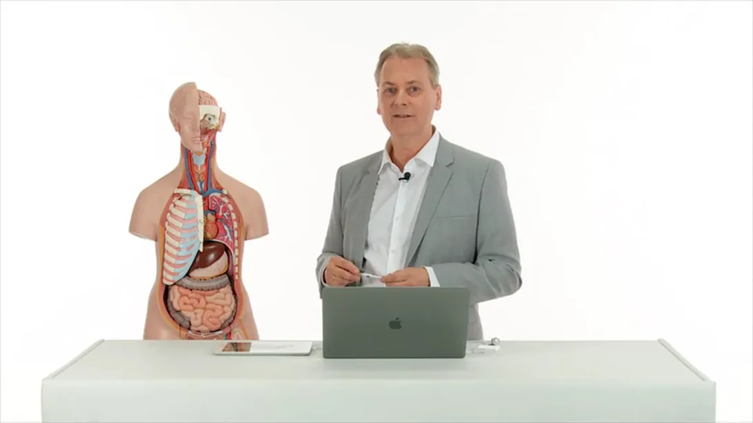

Course Speakers


Curriculum
Watch free lectures
Create a free account or log in to access free lectures for this course.
No payment required.
Create free account Log inChapter 1
Free lectures
1 lesson
Lectures & Quizzes:
Chapter 2
0.5 CME
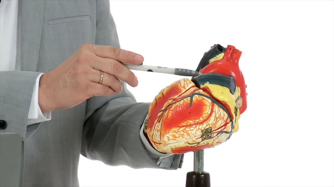
Heart
This chapter is designed to provide you with a focus on sonographic planes, terminology and probe manipulation skills, along with techniques involved in handheld ultrasound Echocardiography to evaluate the heart chambers in 2D. Learn how to differentiate normal heart function from major cardiovascular pathology in this focus tutorial. The professional will also identify situations when a referral for a second opinion is indicated.
1 lesson and 1 quiz
Lectures & Quizzes:
Chapter 3
0.5 CME
Lung I
In a first glance Prof. Thomas Binder will talk about pleural effusions. Next to come are artifacts of the lung such as A-lines and B-lines, how to identify a pulmonary edema and you will learn how you can quickly and accurately identify a pneumothorax.
1 lesson and 1 quiz
Lectures & Quizzes:
Chapter 4
0.5 CME
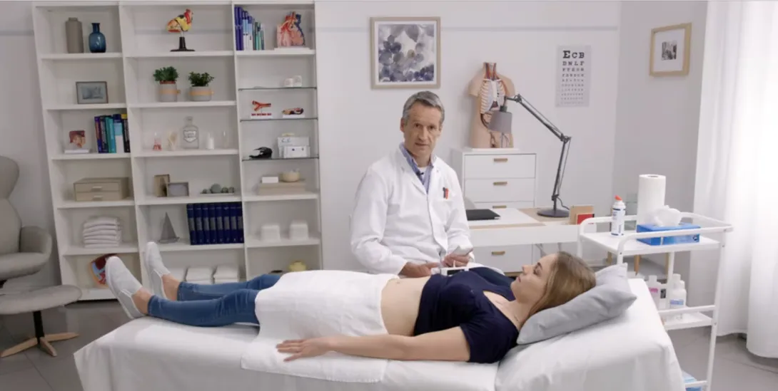
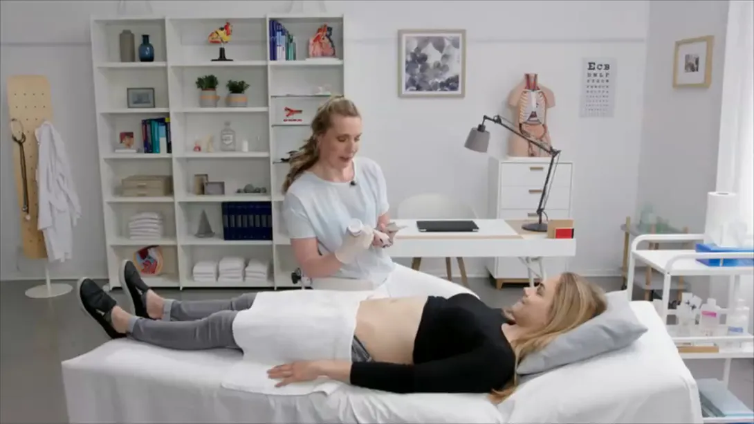
Obstetrics
This chapter is designed for healthcare professionals who have an interest in point of care obstetric ultrasound. The chapter will provide you with probe manipulation techniques and an early pregnancy ultrasound protocol with handheld devices. You will experience how to differentiate ectopic pregnancy and you will learn about the associated probe manipulation technique. The professional will also identify situations when a referral for a second opinion is indicated.
1 lesson and 1 quiz
Lectures & Quizzes:
Chapter 5
0.25 CME
Kidney
This chapter aims to improve your understanding and use of handheld ultrasound to answer clinical questions by focussing on sonographic planes, terminology and probe manipulation skills. The professional will be able to identify the sonographic appearances of normal and abnormal structures required to answer common clinical questions and understand situations when a referral for a second opinion is indicated.
1 lesson and 1 quiz
Lectures & Quizzes:
Chapter 6
0.5 CME
Urinary tract
This chapter aims to improve your understanding and use of handheld ultrasound to answer clinical questions by focussing on sonographic planes, terminology and probe manipulation skills. The professional will be the variants and abnormalities of the urinary tract from presentations, hands-on demos and case-based learning. The professional will be able to understand situations when a referral for a second opinion is indicated.
1 lesson and 1 quiz
Lectures & Quizzes:
Chapter 7
0.25 CME
Biliary tract
This chapter was designed for healthcare professionals who aim to incorporate handheld ultrasound assessment of the biliary tract in their practice. The professional will learn how to recognise the variants and abnormalities of the biliary tract from presentations, hands-on demo and case-based learning and understand situations when a referral for a second opinion is indicated.
1 lesson and 1 quiz
Lectures & Quizzes:
Chapter 8
0.5 CME
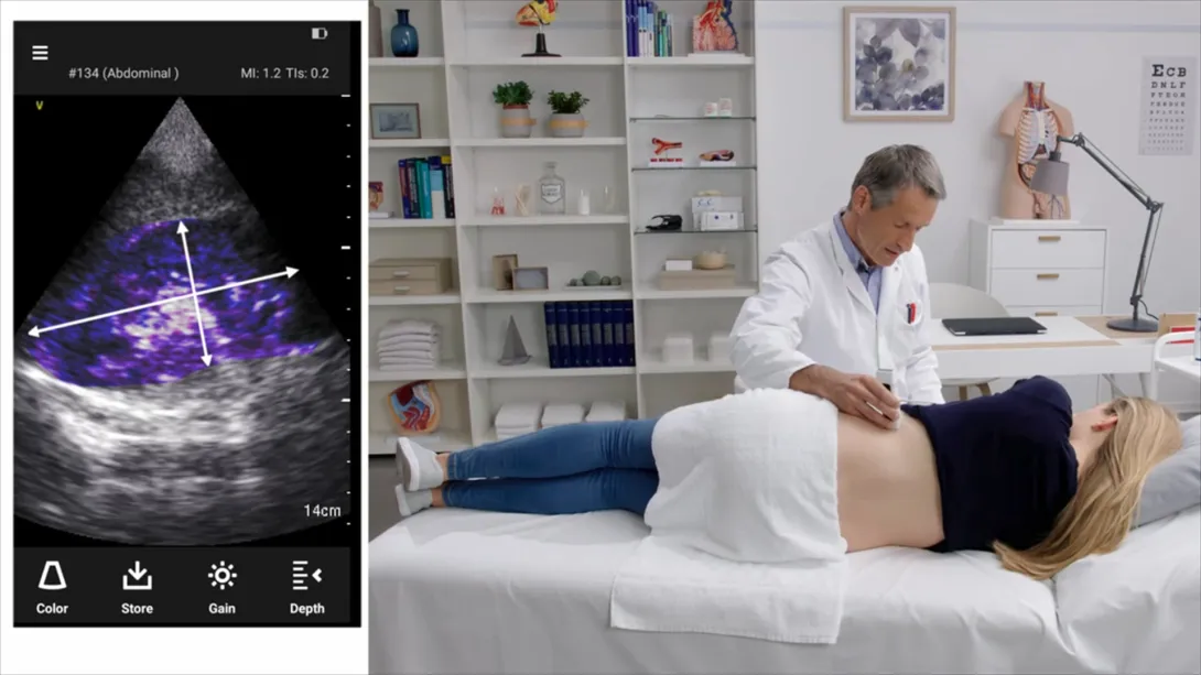
Abdominal Aorta
This chapter aims to improve your understanding and use of handheld ultrasound to answer clinical questions in your practice by focussing on sonographic planes, terminology and probe-manipulation skills. The topic provides an overview of anatomical structures and vessels around the abdominal aorta where the professional will understand variants and abnormalities as well as situations when a referral for a second opinion is indicated.
1 lesson and 1 quiz
Lectures & Quizzes:
Chapter 9
0.75 CME
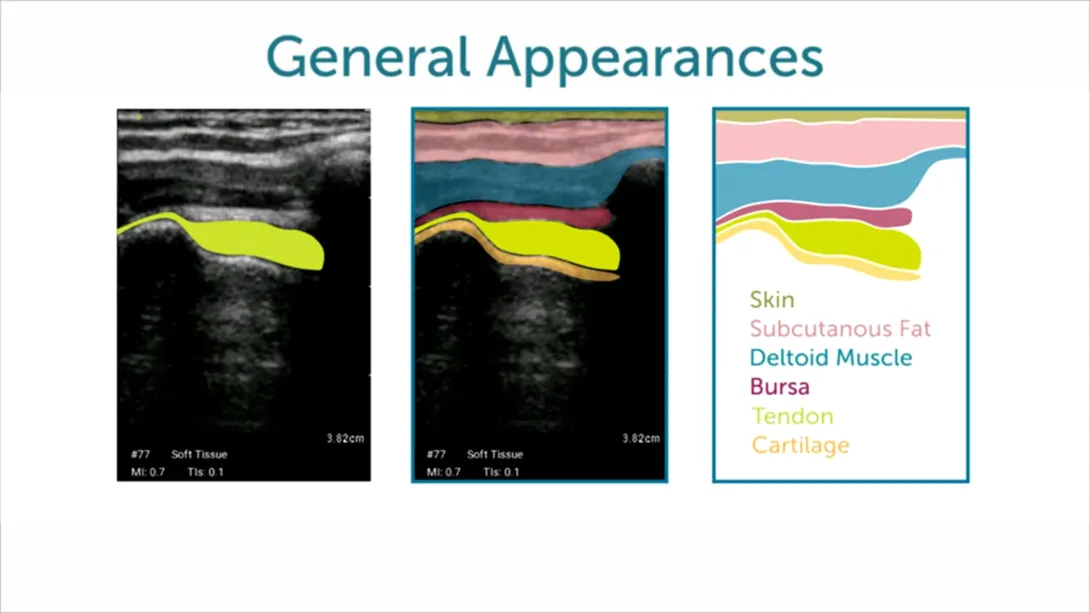
MSK
This chapter provides an introduction to the anatomy and ultrasound examination of the knee and shoulder. Particular emphasis is given to scanning protocols as well as tips and strategies for obtaining high-quality images. The professional will be able to identify the sonographic appearance of normal and abnormal structures required to answer common clinical questions and understand situations when a referral for a second opinion is indicated.
2 lessons and 1 quiz
Lectures & Quizzes:
Chapter 10
0.5 CME
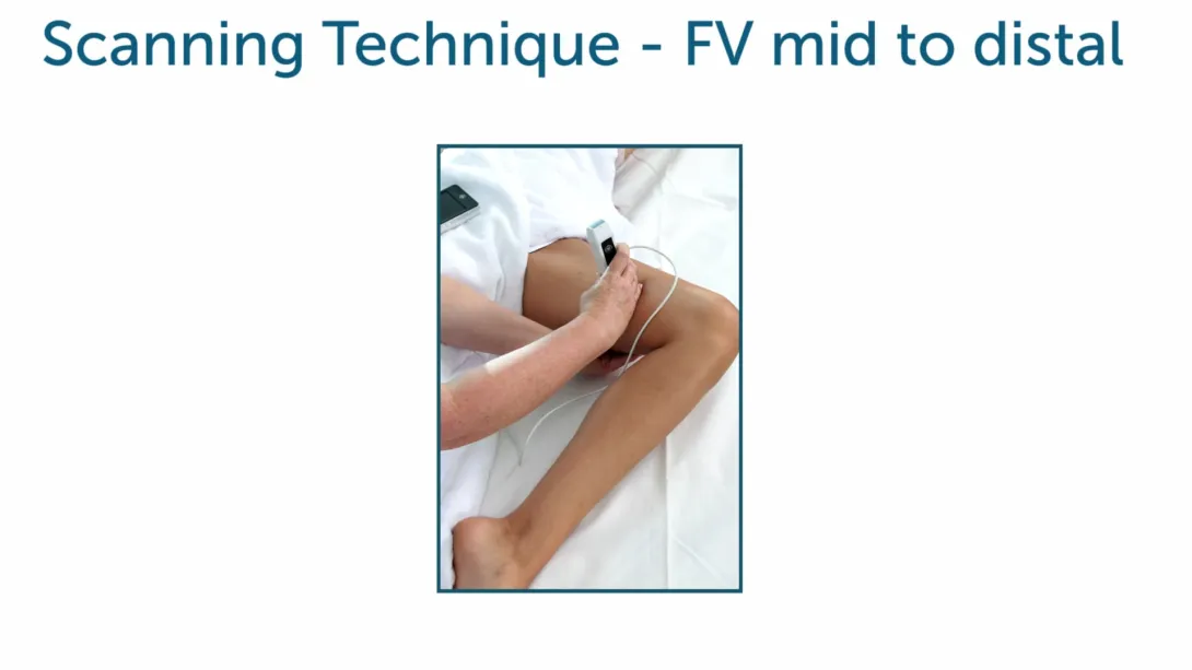
DVT
This chapter aims to improve your understanding and use of handheld ultrasound to answer clinical questions by focussing on sonographic planes, terminology, and probe manipulation skills. Importantly, an overview of anatomical landmarks of the peripheral venous system is included as part of this chapter. The professional will recognise the variants and abnormalities of the peripheral venous system from the presentation of a hands-on demo and case-based learning and will understand situations when a referral for a second opinion is indicated.
1 lesson and 1 quiz
Lectures & Quizzes:
Chapter 11
0.75 CME
Carotid
Imaging the carotid arteries is "straight forward". In this chapter we will explain, which instrumentation you need and how a carotid ultrasound exam is performed. Here you will also review the anatomy of neck arteries and learn when a carotid scan is indicated in a point of care setting. After completion you will know how to assess the Intima Media Thickness (IMD) and detect plaques to assess cardiovascular risk. In addition, we cover the topic of "carotid artery dissection".
1 lesson and 1 quiz
Lectures & Quizzes:
Chapter 12
0.5 CME
eFAST
Here we teach you the eFAST protocol, which is used in the ultrasound assessment of trauma patients. Niko and Martin explain when it should be used and which views you should acquire. Learn which pathologies you can detect and watch teaching cases, which highlight the potential of the eFAST exam.
1 lesson and 1 quiz
Lectures & Quizzes:
Chapter 13
0.5 CME
Lung II
Scanning the lungs is not difficult to learn and it can easily be combined with the ultrasound of other organs. This chapter covers the sonographic anatomy and demonstrates how to image the lungs. We provide you with in-depth insights into pleural effusion, pulmonary congestion, consolidations and pneumothorax.
1 lesson and 1 quiz
Lectures & Quizzes:
Objectives

After completing the Point Of Care Ultrasound FocusClass, you will be ready to successfully apply hand-held ultrasound in your point of care environment.
You will have a broad understanding of instrumentation, ultrasound anatomy and pathology.
Ideal for:

Student Discount
Are you a student? Get 50% discount on this course by completing the student application form.
Get Student DiscountRecommended Blog Posts


Pricing
One-Month Access
Take the most flexible route with a monthly subscription.
You get:
- Cancellation possible anytime after 4 months minimum run time
- Ability to complete quizzes and earn CME credits
Half-Year Access
Our shortest option for very fast learners. Ideal for people who have plenty of time to learn.
You get:
- 6 months access to our course
- Ability to complete quizzes and earn CME credits
One-Year Access
Our most recommended access duration to dive deep into the course. Save 30% on the 6-month option.
You get:
- 12 months access to our course
- Ability to complete quizzes and earn CME credits
Two-Year Access
If you want to take your time learning, this option is perfect for you. Save 40% on the 6-month option.
You get:
- 24 months access to our course
- Ability to complete quizzes and earn CME credits