

Course Speakers


Curriculum
Watch free lectures
Create a free account or log in to access free lectures for this course.
No payment required.
Create free account Log inChapter 1
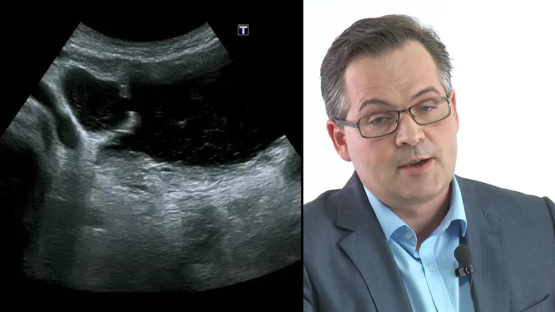
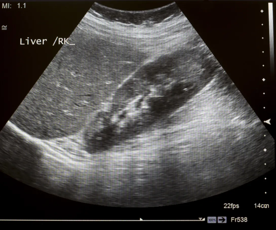
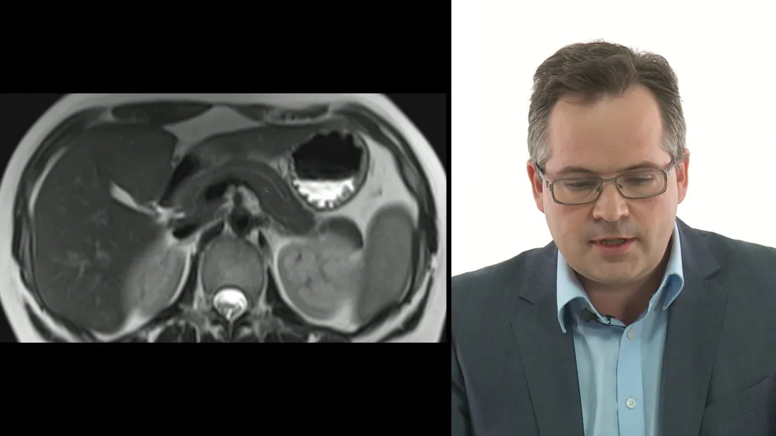
Abdominal Ultrasound
This chapter provides a comprehensive introduction to abdominal ultrasound, covering essential techniques and anatomy for various organs. You will meet the speakers of the Abdominal Ultrasound BachelorClass and explore clinical case reports showcasing the utility of abdominal ultrasound. The main part includes detailed lectures on scanning the liver, gallbladder, kidneys, bladder, spleen, and the pancreas, with practical tips for identifying pathologies and performing thorough exams. More pathology examples can be found in our Abdominal Ultrasound BachelorClass.
11 lessons
Lectures & Quizzes:
Chapter 2
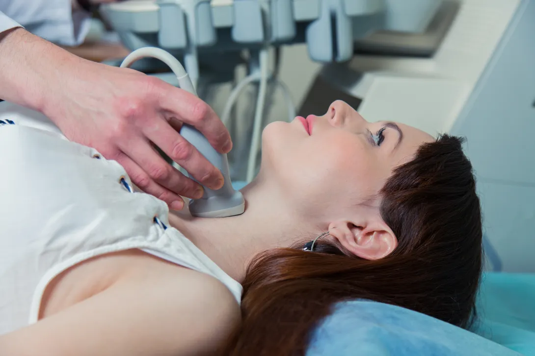
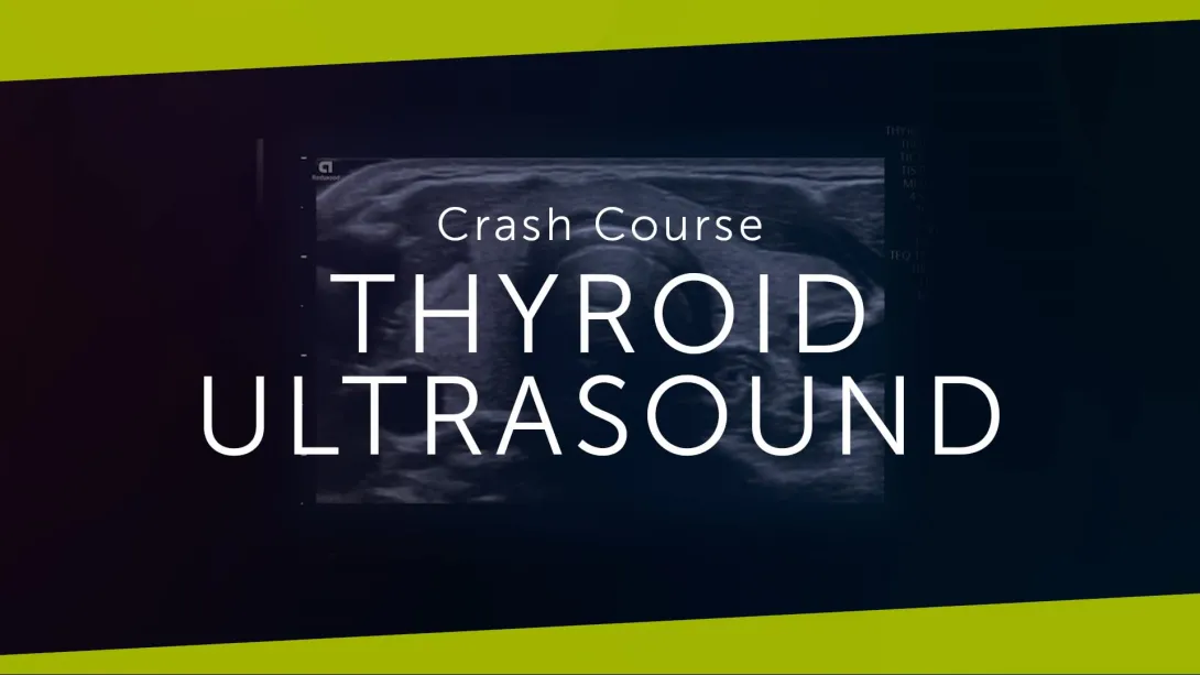
Thyroid Ultrasound
This chapter introduces thyroid ultrasound, covering indications and basic imaging techniques. You will learn how to identify and describe benign thyroid cysts and perform a standard thyroid exam, including patient positioning, image orientation, and ultrasound anatomy. The lectures also cover contour, texture, shape, and size quantification of the thyroid, with many tips on how to image the vessels for additional information.
6 lessons
Lectures & Quizzes:
Chapter 3
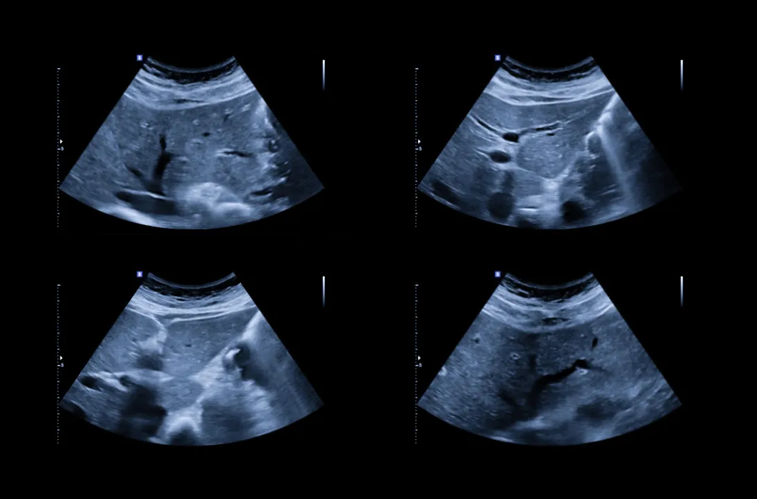
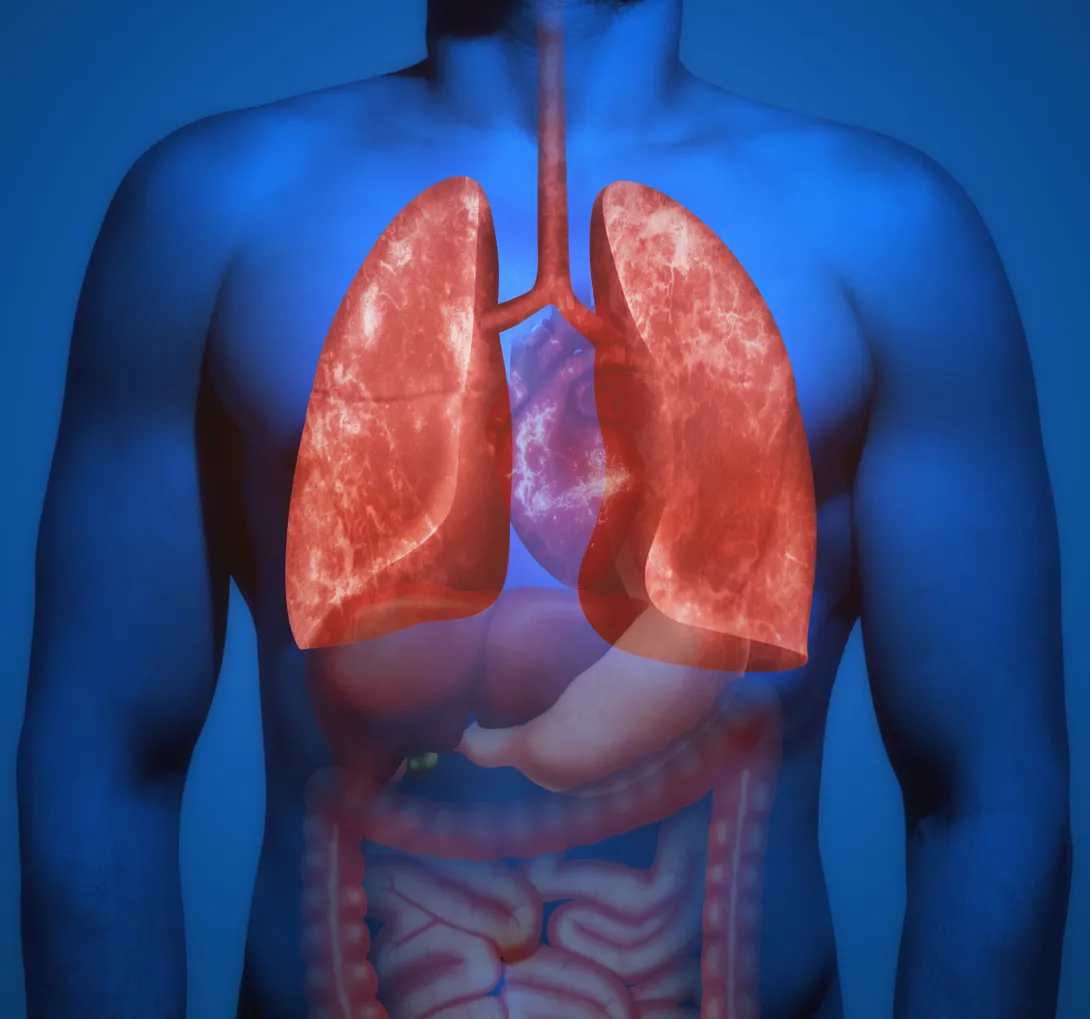
Lung Ultrasound
This chapter on lung ultrasound covers the basics, including pleural effusion, B-lines, pneumothorax, and consolidations. You'll learn to recognize diffuse parenchymal lung disease and use ultrasound for diagnosing COVID-19 viral pneumonia.
5 lessons
Lectures & Quizzes:
Chapter 4
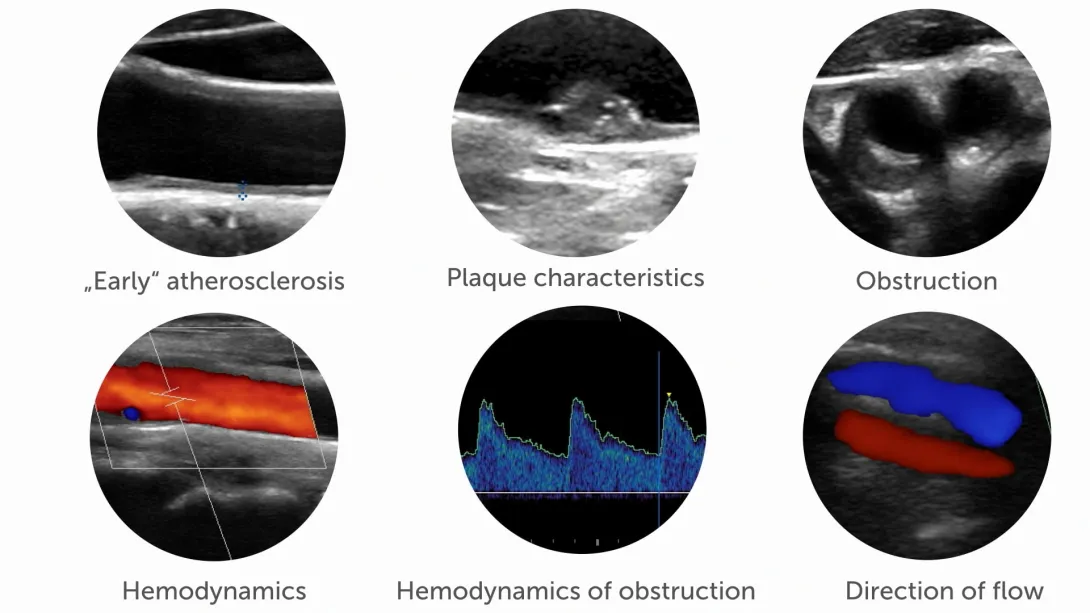
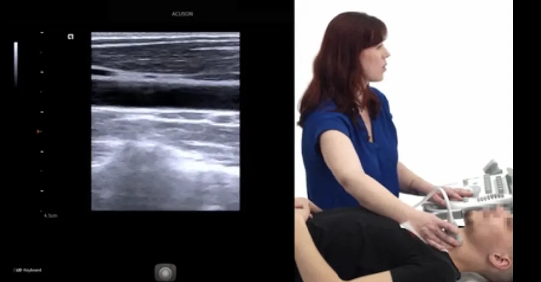
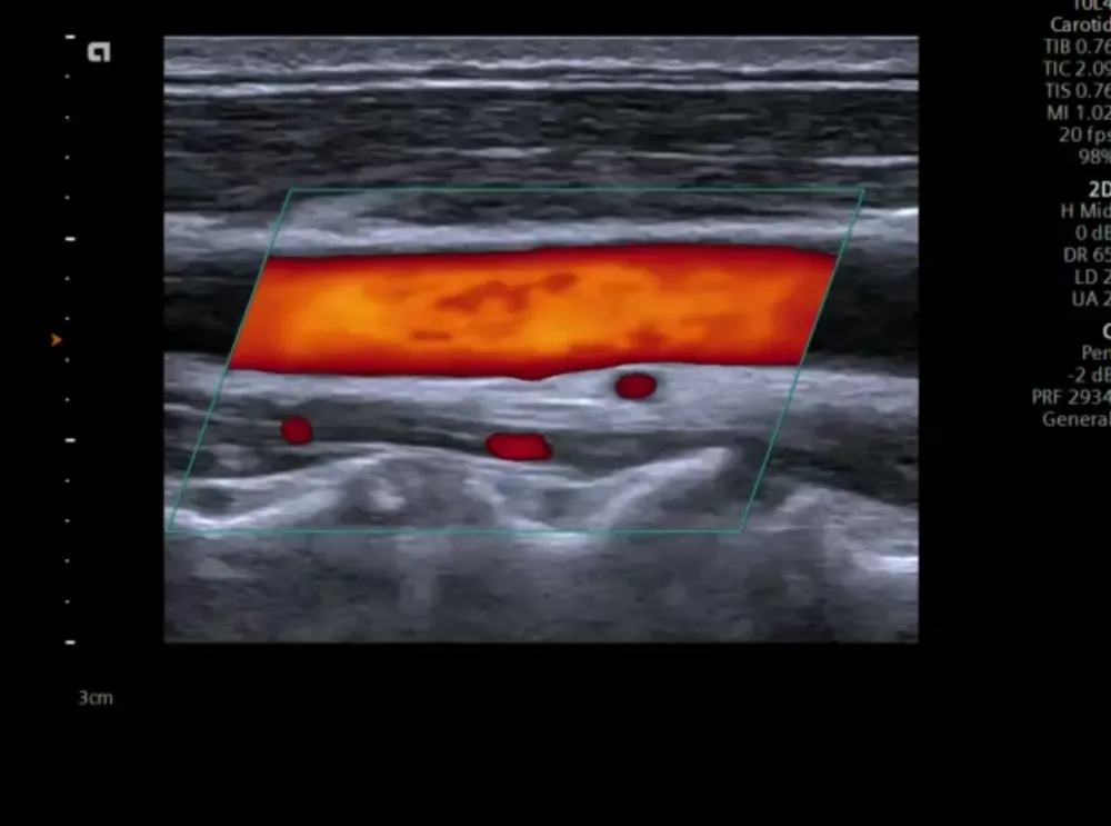
Carotid Ultrasound
This chapter teaces the essential techniques and best practices of carotid ultrasound. You will learn how to obtain the basic imaging planes, with practical tips for challenging cases and ergonomic advice for positioning yourself and the patient. The lectures guide you through scanning the carotid arteries from the proximal section to the most distal parts at the base of the skull, using transverse and longitudinal planes, and include numerous examples in both B-Mode and color Doppler. Additionally, the chapter covers the effective use of color Doppler, addressing blood flow direction settings, optimal angulation, and proper color box settings.
5 lessons
Lectures & Quizzes:
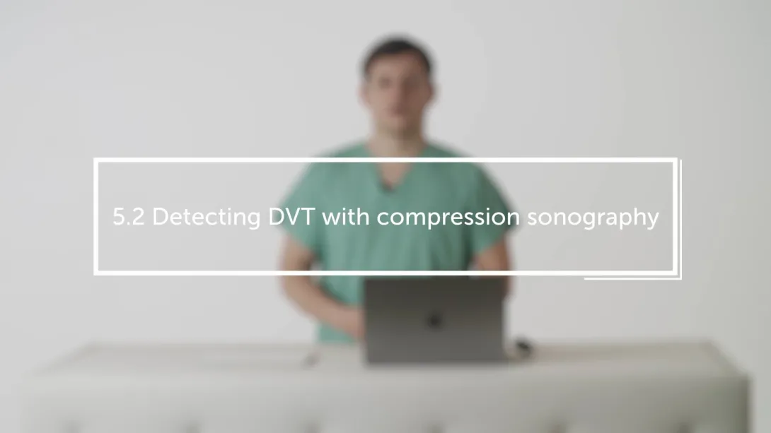
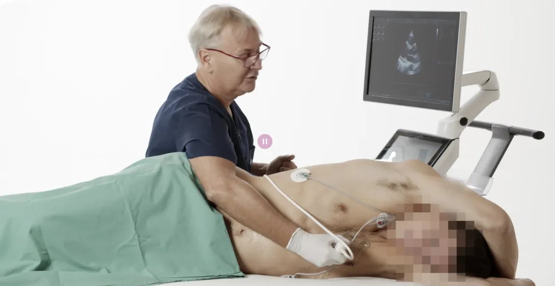
Chapter 5
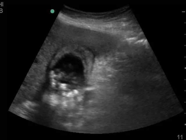
Compression Sonography of the Deep Veins (DVT)
This lecture was originally filmed for our Emergency and Critical Care Ultrasound Essentials - It shows how to quickly and effectively diagnose or rule out deep vein thrombosis of the lower limb! You can learn more about DVT and Pulmonary Embolism in the Emergency and Critical Care Ultrasound Essentials Course!
1 lesson
Lectures & Quizzes:
Chapter 6
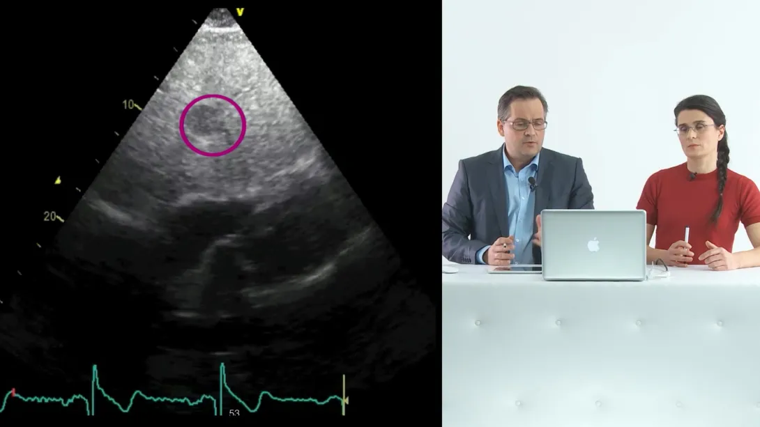
Basics of Cardiac Ultrasound
Imaging the heart presents unique challenges compared to other parts of the body. The lungs and ribs obstruct the view, and the heart itself is constantly in motion. In this chapter, we will explore the specialized instruments and techniques used to overcome these obstacles. You will learn about the various imaging modalities included in a routine cardiac exam and the specific imaging windows applied. We will also cover the standard views essential for a comprehensive assessment. Through detailed, step-by-step guidance, we will demonstrate the process of performing echocardiography, ensuring you gain a thorough understanding of this vital diagnostic tool.
4 lessons
Lectures & Quizzes:
Objectives

Learn all the essentials from abdominal ultrasound, over thyroid ultrasound to carotid and DVT scanning and even the basics of echocardiography.
Learn how to image all radiology-relevant body regions.
Increase your confidence and practical imaging skills.
Ideal for:

Student Discount
Are you a student? Get 50% discount on this course by completing the student application form.
Get Student DiscountRecommended Blog Posts
Pricing
One-Month Access
Take the most flexible route with a monthly subscription.
You get:
- Cancellation possible anytime
- Lectures, micro- and nano-learning
One-Year Access
Billed yearly.
You get:
- 13% savings on yearly plan
- Lectures, micro- and nano-learning