2. Carotid Ultrasound - Anatomy

2.1 Which structures do we see on carotid ultrasound?
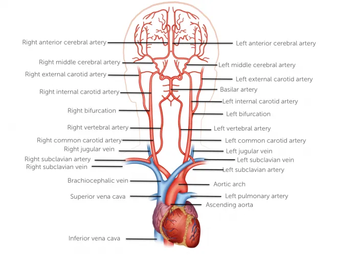
Carotid ultrasound allows you to display the extra cranial vessels of the neck. This includes the carotid arteries and its branches: the internal and external carotid arteries and the vertebral arteries. In addition, we can also see the jugular veins, in particular the internal jugular vein. Carotid (Duplex) ultrasound also displays non- vascular structures of the neck such as the thyroid, lymph nodes, thyroid cartilage, muscles of the neck and nerves. It is equally important to identify these structures since they not only serve as anatomical landmarks but can be involved in pathologies. Such pathologies should be picked up during a carotid Duplex ultrasound exam as well.
2.2 What are some variations of anatomy that you need to be aware of ?
- The level of the bifurcation can vary
- The size and location (anterior/posterior, medial lateral) of the internal and external artery is highly variable
- Shape of the bifurcation (angle of exit of the internal and external carotid artery)
- Origin the right and left common carotid artery can vary
- Size of the vertebral artery (aplasia, hypoplasia)
- Origin of the superior thyroid arteries from the common carotid artery
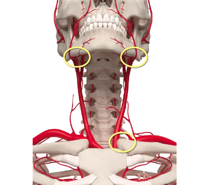
2.3 Which regions of the carotid artery are difficult to image?
| Difficult regions to image |
|---|
| Cranial portions of the external and internal carotid arteries |
| Origin of the left common carotid artery |
| Origin of the vertebral arteries |
| Cranial segments of the vertebral arteries |
| Segments of the vertebral arteries that are posterior to the transverse processes |
2.4 What are the branches of the common carotid artery?
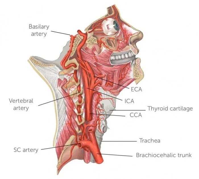
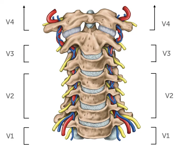
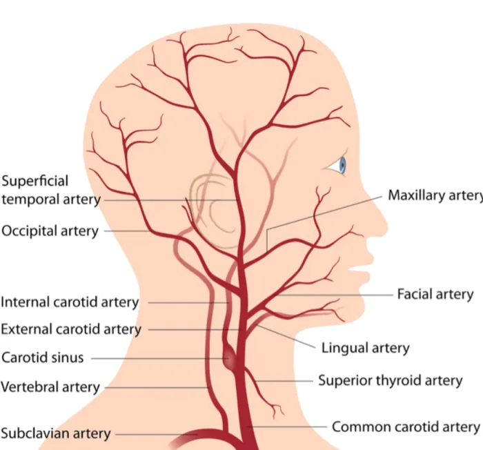
The right common carotid artery originates from the brachiocephalic trunk, while the left common carotid artery arises from the aortic arch. The common carotid artery separates into the internal and external carotid artery at the upper boarder of the thyroid. This region is also called bifurcation. The internal carotid artery enters the skull through the carotid canal where it bifurcates into the middle cerebral artery and anterior cerebral artery forming the circle of Willis. The vertebral artery supplies the spinal chord and the cerebellum with blood. It arise form the subclavian artery and has 4 segments: The pre-foraminal segment, the formaminal segment (which runs in the transverse foramina of C6-C2), extradural segment, intradural segment.
2.5 What are the branches of the external carotid artery?
In contrast to the internal carotid artery the external carotid artery has 8 extracranial branches. These are:
- Superior thyroid artery
- Ascending pharyngeal artery
- Lingual artery
- Facial artery
- Occipital artery
- Posterior auricular
- Maxillary artery
- Superficial temporal artery
2.6 What are the layers of the artery wall?
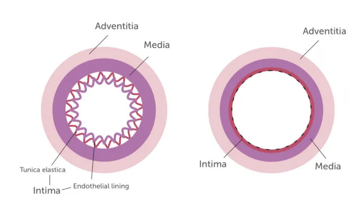
A very important part of carotid ultrasound is the assessment of the intima media thickness (IMT). The intima media is easy to discern with ultrasound. The intima is typically bright (echogenic), while the media is darker (hyoechogenic) than the intima and the adventitia. The intima media thickness has been used to assess the risk for cerebrovacualar disease and to estimate “vascular age”. Measurements of intima media thickness should always be performed during a carotid (Duplex) ultrasound exam.
Intima: Inner layer, consists of an endothelial lining of endothelial cells and an elastic membrane (tunica elastica)
Media: Middle layer, muscular and elastic components
Adventitia: Collagen tissue, contains vasa vasorum (in larger arteries)
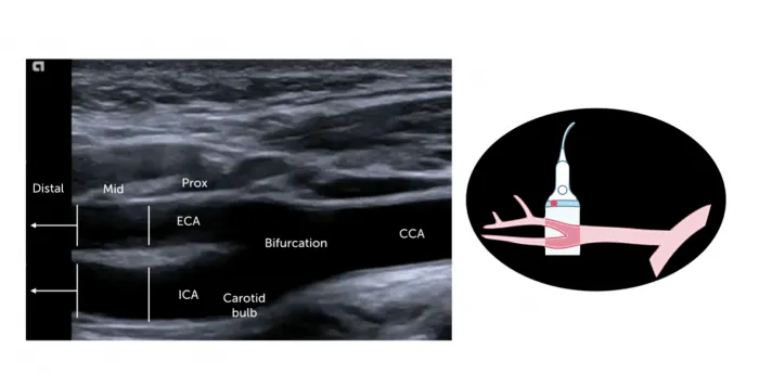
2.7 What are the segments of the carotid artery in ultrasound?
The carotid artery can be divided into the following segments:
- Common carotid artery
- Proximal (origin from the subclavian artery or brachiocephalic trunk)
- Mid
- Distal (below the bifurcation)
- The Bifurcation (which also includes the proximal portion of the carotid bulb)
- Internal carotid artery
- Proximal (which includes the distal carotid bulb)
- Mid
- distal
- External carotid artery
- Proximal
- Mid
- Distal
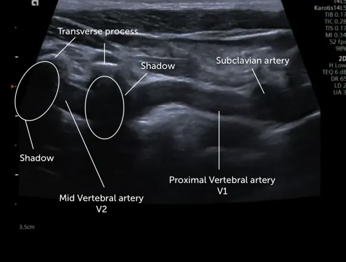
2.8 How do I identify the vertebral artery with ultrasound?
The vertebral artery is located posterior and medial in relationship to the common carotid artery. The proximal portion (origin from the subclavian artery) is often difficult to visualize, especially in elderly patients. The vessel has a more or less tortuous course and is interrupted on ultrasound due to shadowing from the transverse processes.