Can you judge which of the left atria shown in our case is visually enlarged?
Case stories that stick
Let’s look at a patient's case that nicely demonstrates the most common symptoms and typical echo features of patients with amyloid heart disease.
A 37 yo woman comes to the emergency department at 4 am with massive cramps in her lower abdomen. The gynecologist on duty performed a transvaginal ultrasound – Let’s have a look!
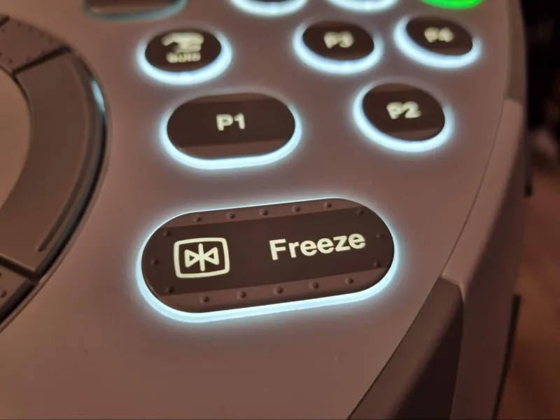
In this part, we have prepared a short video for you about documentation and quantification in ultrasound because, after all, measuring and saving our findings is an essential part of our work as sonographers.
In part 8 of the Ultrasound 101-series, we will learn about why not all things on your scanner’s image are as they seem.
Here’s a case from our Adult Congenital Heart Disease BachelorClass: A 60 y/o woman with palpitations and no relevant previous medical history presents at your echocardiography lab.