
Writing an Ultrasound Report – Secrets: Part 4 – 6 Tips that will help you design and structure your report


As health care professionals we are mainly concerned about the content and phrasing of our report. But even the best medical report needs a good framing. This is where “design” and structure of a report come into play. Here are my 6 tips that will make your report shine:

1) „Design“ your report.
Steve Jobs once said:
“The design is not just what it looks like and feels like. The design is how it works”
This holds true also for a written medical report. Aside from the fact that it brands your institution, clinic or you as a person a good design also helps the reader to quickly extract the relevant information.
In a world where time is extremely valuable we often browse more than we read. So use graphic elements such as lines, arrows or boxes and color to make clear what belongs to what and highlight the relevant information. It is equally important to use a font that is readable, so don’t pick anything fancy and playful such as the following:
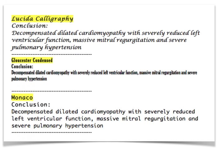 Example of fonts, which are difficult to read and should be avoided.
Stick to fonts such as: Helvetica, Georgia, Verdana or Cambria. These fonts are among the easiest to read and also symbolize trust and authority.
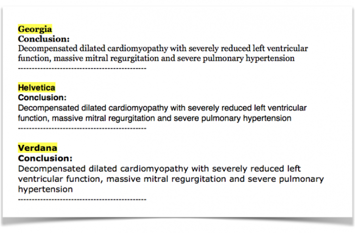 Example of fonts, which are easy to read and symbolize “authority”.
Obviously there is more to design than fonts, graphic elements and a logo. So depending on your budget and goals it might even pay off to consult a professional design expert.
2) Patient info: Highlight what is important:
Put yourself into the position of a physician who reads your report. What does he need to know first - aside from the patient name and date-of birth? The hospital administration number or the name of the referring physician? Or is it the quality of the study?
None of these: clearly it is the type of study (i.e. “Ultrasound of the Abdomen”) and the date of the exam!
Unfortunately these valuable pieces of information are often hidden somewhere in the report. Obviously you want to know “what report you are confronted” with. It also makes a big difference if the report is 1 day, a week or 5 years old; Especially, if patients come to your office with a huge portfolio of medical reports and you have to sort them along a timeline.
So here is my suggestion: Highlight the following 1) type of exam, 2) name, date of the patient 3) date of birth, and 4) the date of the exam on your report. Everything else such as administration numbers, time of referral, referring physician, social security number, the phase of the moon or anything else can be included in a less prominent section of the report.
3) Write a conclusion
Many physicians (including myself) often read the report from the bottom to the top, starting with the conclusion. We need to first get an overview before we dig into the details. Unfortunately many reports do not include a conclusion or do not provide the information I am looking for. Here is my suggestion: Provide a clear conclusion section at the end of the report, which stands out. State the most relevant findings and its interpretation there. Here is an example:
 Example of a conclusion, which provides an interpretation of the findings and a recommendation for further evaluation.
4) Structure the main text
Discipline yourself to always use the same sequence of describing your findings. In abdominal ultrasound you might want to start with the liver then the gallbladder followed by the pancreas. In echocardiography I always start with the ventricles (left then right) then the atria followed by the valves etc. I also recommend to always use the same sequence when describing a certain organ or structure. For example: “Normal size, shape, surface and structure of the liver”. Not only will the referring physician know that these features were “looked at” but it also prevents you from writing an incomplete report.
5) Include measurements
Quantification is important for many reasons: they are used to make a diagnosis, allow us to detect regression or progression of disease and they might trigger a certain form of therapy or intervention. Therefore it is important to include a table with all relevant measurements. While this has become common practice in many labs I would also recommend to add the normal ranges, use measurements, which are indexed to body surface area and to also include important values, in the text body of your report. This way the measurements do not have to be looked up in the tables but stand out.
6) Add Images
“A picture says more than a thousand words”. Adding images is a great way to communicate information. It is also a good way to “show cast” your professionalism and skills.
Images can also be used in an echo report. While you cannot display functional information such as left ventricular function, still image are never the less a good way to give the reader an impression of the problem. For example, the size of the chambers in a 4 chamber view, the morphology of the valve or the shape of a Doppler spectrum. Several structured reporting systems allow you to select images that are included in your report. If you do not have this feature, simply print out some images from your scanner.
True, you will need to put some effort into creating a good report, but it pays off - for you and your patients.
In the next posting we will discuss the topic of “phrasing”. Here we will provide you with some sample reports that you can use as a template.
Best Thomas Binder and the 123sonography team
PS: If you missed the previous topics on reporting here they are:
- Part 1 How an ultrasound report helps your career
- Part 2 Mastering the technology
- Part 3 Biggest mistakes