Writing an Ultrasound Report – Secrets: Part 2 Mastering the technology
Wouldn’t it be great if your ultrasound report would generate itself? With the click of a button all the images and measurements are analyzed, interpreted and the findings are translated into text. All you need to do is press “print”.
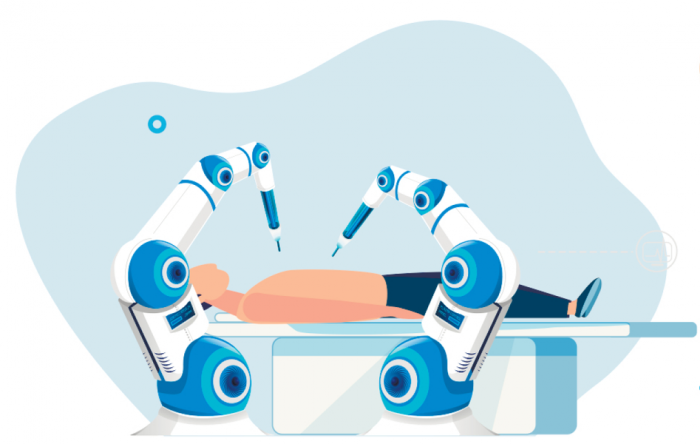
 Artificial intelligence is already affecting the workflow in ultrasound.
Artificial Intelligence and workflow
While such a scenario is still science fiction, there is a huge push in this direction. I am convinced that “artificial intelligence” will not only affect the way health care professionals will practice ultrasound but also help us to generate a report.
But where are we now? How can new technologies such as artificial intelligence, speech recognition and other IT technologies assist with reporting?
Newer scanners already incorporate AI driven technologies to enhance workflow. Here are some examples:
- Automatic image optimization – The scanner continuously adapts the setting to ensure that you always have the best possible image quality. No matter if you are using B-Mode or spectral Doppler.
- Automatic measurements – Doppler curves are automatically traced and measured. The values are displayed on the screen with no need to even touch the trackball. A great way to standardize your measurements!
- Automated endocardial- and IMT tracking. The 2D image is analyzed to detect boarders and boundaries on the image. This allows all sorts of automatic calculations in cardiology and vascular ultrasound such as chamber volumes or Intima Media Thickness (IMT)
- Machine learning algorithms learn from image datasets to make “predictions”. This discipline of artificial intelligence is used, for example to recognize views, tissue characterization, segmentation and the detection of tumors.
Reporting: “Two-point-zero”
These are only a few of the many developments that help us to generate and analyze ultrasound images. But will new technologies also help us to generate a report? What do we currently have at hand?
 Digital technology is increasingly used to generate reports. Such reports can easily integrated into the digital medical record and viewed from mobile devices.
One technology that is increasingly used in radiology is medical speech recognition. Such systems automatically transcribe your dictation into written text. While older systems were difficult to use and not very precise, new software is quicker and superior in understanding medical terms and phrases. Your text is generated 3-5x faster than by conventional type writing and there is no need to wait until your secretary transcribes your dictation. Most speech recognition solutions also permit “voice navigation” so that you can quickly make corrections and give commands to operate the software.
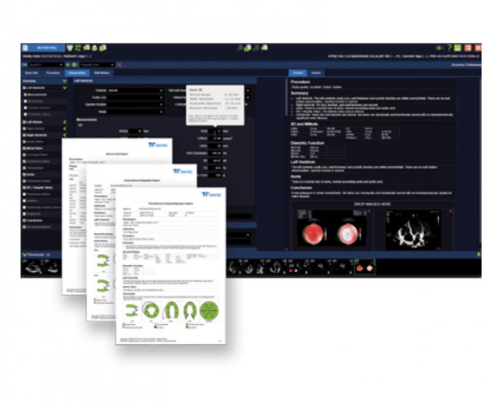
The second methodology you can use is “structured reporting”. There are several vendors including ultrasound providers, which offer text block solutions to generate a report. In essence all you need to do is go through a check box worksheet that lists specific classifications of commonly described structures and features. You can also grade specific findings (i.e. severity of enlargement). Individual text blocks are added to the report for each selection you make. Since the reporting software is (or can be) connected to your scanner, measurements are automatically transferred into the report. You can also select images that should be included in the printout of the report.
 Step by step approach to reporting using a structured worksheet. Measurements (including normal values) and graphics can be included.
Adhering to guidelines
Structured reporting has a major advantage - especially for ultrasound novices: The definitions and classifications, which are applied, correspond to the newest guidelines and you do not have to worry about phrasing. Since the report is “structured” it reminds you of what should be included in a report. The templates can usually be adapted to the specific needs of the user and it is possible to integrate graphics which help to communicate your findings to the referring physician.
Is it worth upgrading to such solutions?
The clear answer here is yes, or at least to move in this direction. Better to generate a word document than a handwritten report. Most institutions already provide infrastructure that you can use. If you don’t have an institution to back you up you might even develop your own system. Many Database Management Systems (DMS) are easy to develope. With a little bit of help you might even program the system yourself. This is actually how we started 30 years ago at the Medical University of Vienna. Our system has now grown into a flexible structured reporting system and database, which allows us to generate a detailed report within minutes so that it is ready often before the patient leaves the lab. A search algorithm also permits us to analyze the data for research purposes.
So while, effort is necessary in the beginning, the pay off in the end is huge.
What is your experience? Which system do you use? I would be curious to know!
PS: In part 3 of the teaching series on “Reporting in Ultrasound” we will look at common mistakes and discuss how you can avoid them.


