
Turn Lights On

Echocardiography is usually performed in the dark, but sometimes it makes sense to turn the lights on. You might not be able to see the echo screen so well but it will provide a glimpse at the patient. Let me tell you a story to show you what I mean....
Just the other day, I examined a patient referred from our nephrologists with shortness of breath. She was a skinny and fragile (152cm, 46kg) elderly lady with a history of kidney failure following polycystic kidney disease. A year ago she underwent kidney transplantation and was doing quite well until she had an episode of acute transplant rejection two months later. Fortunately, this was recognized early and she recovered quickly after treatment.
Anything that disturbs you on this echo?
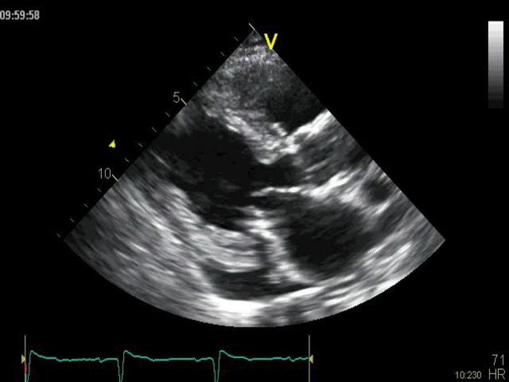
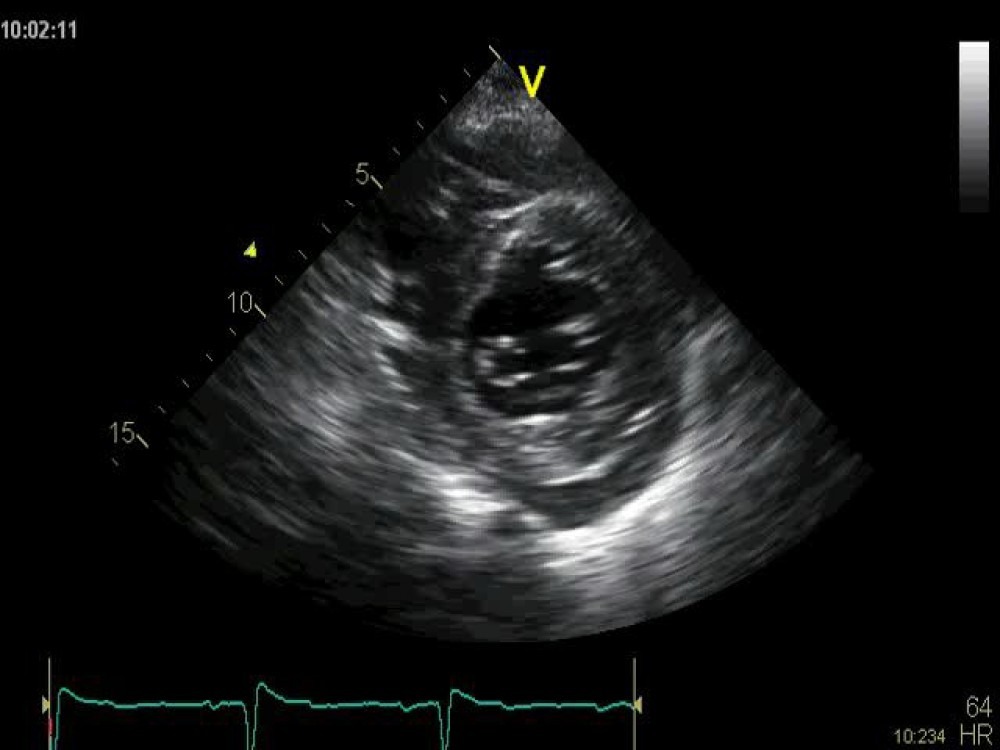
Well here is her echocardiogram, there is something that I would like to point your attention to. Take a look first and see if you can find something that might be important for the management of the patient:
Let me walk you through...
It is not unexpected that the patient has left ventricular hypertrophy - after all, she has kidney disease and thus a long history of hypertension. Yes, she also has a pericardial effusion. But that too is not unusual in such patients. Clearly, the effusion is insignificant and cannot explain her symptoms. Mitral annular calcification is also common in such patients. With the exception of mild mitral regurgitation, the valves are functioning well.
What about the ventricle?
Don‘t you think it is rather large for such a small patient? The end-diastolic diameter was 45mm and the end-diastolic volume 80 ml. Remember she is a small lady (body surface area = 1.34). These values clearly denote dilatation when they are indexed to her body surface area (59). The upper limit of normal for the end-diastolic volume is 50.
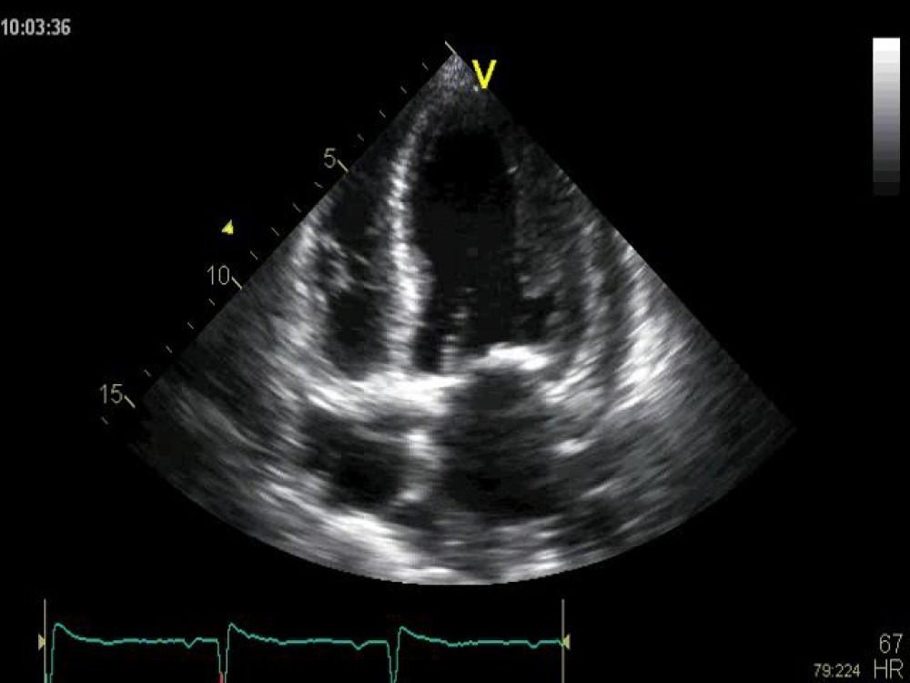
And what about left ventricular function? Let us have a second look:
Left ventricular function is certainly not reduced. As a matter of fact, it is hyperdyamic. The ejection fraction was 78%.
What does this mean?
A dilated left ventricle with hyperdynamic function means volume overload. All we now need to do is find its cause: We have already excluded mitral and aortic regurgitation which are the most common reasons for volume overload. A patent ductus arteriosus (PDA) was also not present. So what else could be the reason?
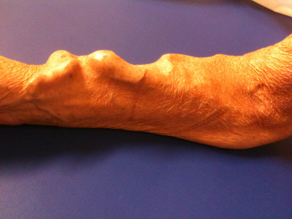
Now it was time to turn the lights on...
She was a kidney patient, right? So what do such patients frequently have? A Cimino (arteriovenous) shunt which is used for dialysis. Here is the image of her left arm.
 Figure 5
Especially if such an ateriovenous shunt is large, as it appears from the image, it can cause significant ventricular volume overload of the ventricle, which eventually can lead to high output failure.
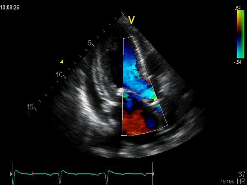
To document the high output state, here is the pulsed wave Doppler signal of the left ventricular outflow tract. The velocity time integral is fairly high (36cm). From this we calculated a cardiac output of 6,5l/min. which is fairly high for such a small patient. By examining the Cimino shunt with Duplex Doppler we calculated a fistula flow of 980ml/min.
 Figure 6
Putting it all together...
This patient clearly had volume overload caused by the Cimino shunt. This probably also explains her symptoms. What is the lesson we have learned?
There are three stumbling blocks on the way to making the diagnosis in this patient:
- First: you have to index the size of the ventricle to the body surface area, otherwise you would not have noticed that the ventricle is dilated.
- Second: you have to know that this could mean volume overload; and
- Third: turn the lights on, sometimes the diagnosis can be confirmed by looking at the patient.
What about the patient? Well, she had the Cimino shunt despite the fact that her kidney transplant was doing fine. The shunt was only left for security reasons since she had a brief episode of rejection. But now she was not on dialysis any more. So we simply suggested closure of the shunt. This should not only reduce the size of the left ventricle but also help to reduce the degree of left ventricular hypertrophy.
If you want to become a true echo expert and see things clearly when others are in the dark, then check out our award-winning Masterclass, which is currently open for registration.
Best,
Tommy