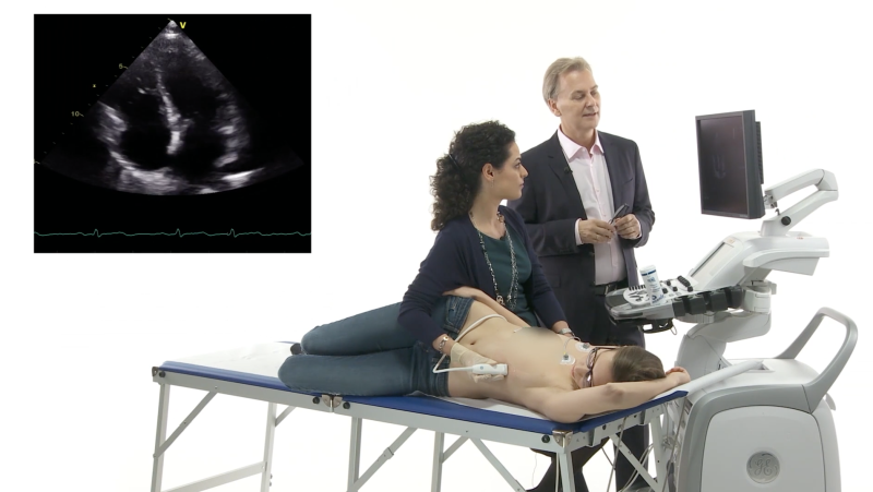
Echo Genius - Image Documentation
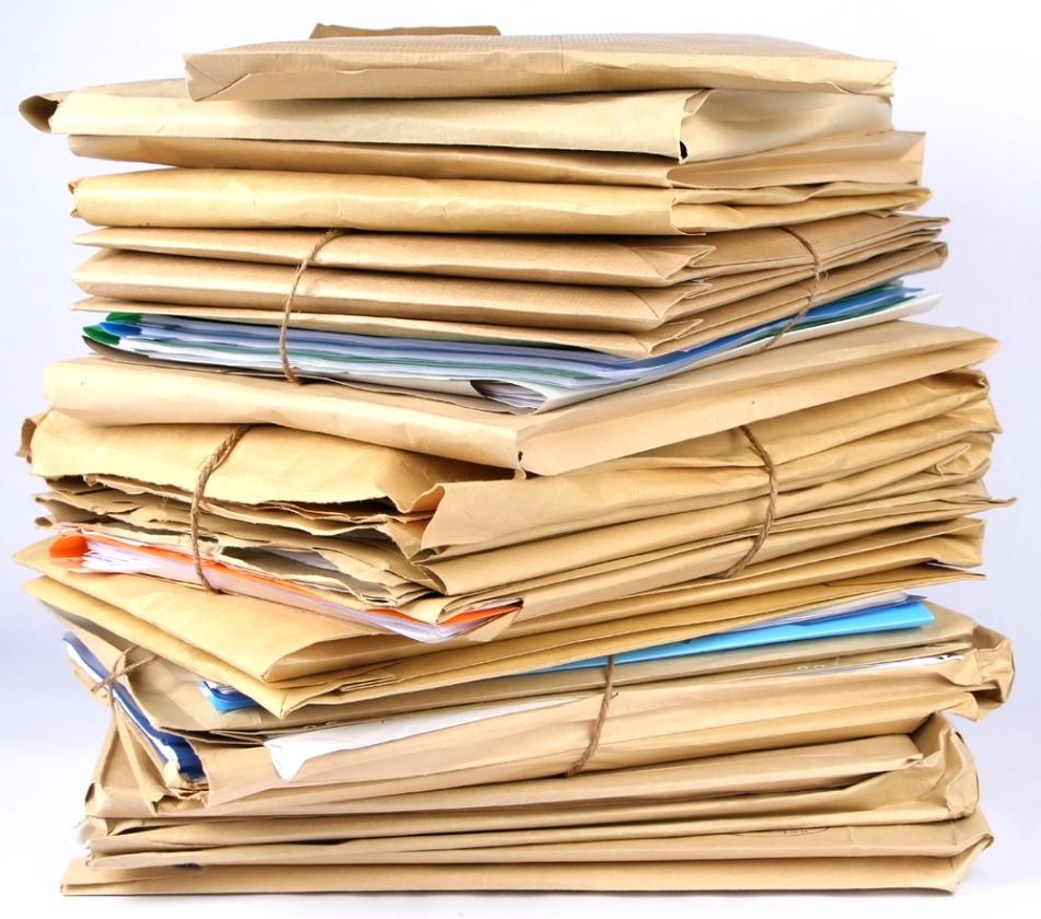
Some colleagues are rather lazy when it comes to documentation of echo exams. However, wouldn’t you agree that taking photos when you go on vacation is a good idea? That having photocopies of important documents can come in handy? Don’t you assume that a copy of your birth certificate is kept somewhere on file? These are just analogs, the point we are trying to convey is: document your exams, it will pay off a thousand times!
There are many reasons why you should document echo exams:
- You can review the studies at a later time and compare them with previous exams. (That’s what radiologists do all the time!)
- You need them to prove what you’ve seen for legal purposes, or to discuss results with colleagues.
- You can perform all sorts of offline analyses (i.e. strain, tissue Doppler, ejection fraction measurements).
- You can easily transfer images to other media or send them to colleagues (or give them to the patient).
- You can use the studies for teaching purposes. (Actually, our own online course would not have been possible without proper documentation!)
And one other important issue: it forces you to perform a complete exam!
But how should you document?
Previously, this was done by recording the exam onto a videotape. While some scanners still have video recorders attached to them, this is a technology of the past. Actually, video recorders are not produced anymore and it becomes more and more difficult to find spare parts to repair them.
Modern documentation is digital! All new scanners allow you to record so-called “image loops”, which display what you record in an endless loop format and present them in some kind of image review library.
To get the best results, it is strongly advisable to use an ECG recording that triggers the start and end of the image loop. This way, the image loops run smoothly, displaying the sequence correctly. Looking at these loops, one gets the impression of a continuously beating heart. If no ECG is recorded, a timer is used (usually 1 or 2 seconds) that defines the length of the loop. These loops are harder to look at without a smooth alignment of cardiac cycles.
We would recommend that you set the machine to record three consecutive beats. That way you even out variations that can occur between beats (i.e., in atrial fibrillation). For contrast studies you might need to record as much as 20 beats (in this case you can also set the recording to a fixed duration of 10 – 20 seconds).
Always review what you record before you store the loop!
You will also need to store still images (MMode, Doppler and still frames where you performed measurements such as the size of a mass, etc.). But it is insufficient to only store still images. After all, you are unable to document functional information on a still image.
A complete exam requires at least 20-25 image loops or still images to be acquired. Implement a standard protocol that defines which views should be recorded. To find the standard protocol for recording echocardiographic views, you can refer to the guidelines provided by the American Society of Echocardiography (ASE). They have detailed protocols for performing comprehensive transthoracic echocardiographic examinations.
This document outlines the recommended views, technical performance standards, and best practices for a comprehensive echocardiographic examination. For additional resources and specific protocols, the ASE website offers various guides and templates (ASEcho) (ASEcho).
What about storage space?
Storage space is no longer an issue. Usually, a complete study consists of 50 to 250 MB, but some studies might be as large as 2GB. But if your hard drive is full, you can always transfer the studies to a central server, which also has the advantage that you can review the studies from other workstations.
Store for comparison purposes!
Do you truly want to provide important, additional, valuable information to the referring physician? Then just do what radiologists do: compare your findings to previous studies! This is easy with digital echocardiography. You can even review loops of different exams, side by side. That way you can pick up even subtle differences such as the size of vegetations or a pericardial effusion of the left ventricular function. After all, the eye is often better than measurements!
What about image formats?
Vendors usually store the images in their own format, which allows all sorts of post processing (raw data). Some use compression algorithms such as MPEG or JPEG, which lead to a minor insignificant reduction in image quality. But all companies must also adhere to a standard image format, which is called DICOM. This allows you to exchange images between platforms, and to read studies with a DICOM viewer.
DICOM also stores study headers that include patient information and measurements.
You can find more on the DICOM format here.
http://medical.nema.org/
You can also transfer and export the images in standard formats, such as MPEG, AVI files, to use them in presentations.
But digital imaging also has a few drawbacks:
- It limits documentation to a few cycles. Therefore, you have to be precise in recording what is important!
- Digital technology is not free of flaws! A hard drive crash can wipe out years of work! So please implement a backup strategy (i.e., RAID system or similar).
- If your server or networks are down, it can easily paralyze your work-flow. In this case, store the images locally and transfer the data later.
- Despite the DICOM standard it can still be a problem to run a network in which scanners from different companies are integrated.
One final word: You will be judged, not only by the quality of your report, but also by how well you document (Just like a photographer)! But that’s okay; it will only force you to try harder and make you a real expert!
In one of our next newsletters, we will talk about the second important component of “quality assurance” – how to write a report!
Recommended articles:
Echo Genius - Writing the Report (Part 1)
Echo Genius - Writing the Report (Part 2)
7 golden rules of echocardiography
