Anatomy is the Key
Hippocrates, Herophilus, Galen, Mondino de Liuzzi, Leonardo da Vinci, Vesalius, William Harvey - what do these names have in common? All of them are famous for their contribution to medicine. And all of them were concerned with human anatomy.

Anatomy is still important - not merely for surgeons, but for those who practice echocardiography as well. I hope the following case will illustrate this fact.
Does the patient have dissection?
A 47-year old patient with hypertension was examined for aortic regurgitation. We obtained a suprasternal view to see whether retrograde flow was present. Look what we found:
 Suprasternal view of the aortic arch. Note: There is an echo-
free structure cranial to the arch, which is separated from the
arch by a “membrane”. Is this an intima flap?
Does the patient have aortic dissection?
 Color-Doppler shows that there is some flow within this
structure. Could it be a false lumen?
The next step:
Obviously, the consequences would be significant because this patient would have to undergo surgery. True, many of you would now perform a CT, which is certainly a good test to rule out dissection. However, there is an easier and more practical way of finding out whether the patient has dissection or not: contrast echocardiography.
We used a contrast agent that is able to pass through the pulmonary vascular system and thus permits opacification of left heart chambers and the arterial system. Look what happened when we injected contrast into a left brachial vein:
 Early phase of contrast application: Note that the structure is
immediately filled with contrast medium.
 Late phase of contrast injection: Now that the aortic arch is
also filled with contrast.
Anatomy revisited
What does this mean? First: the structure cranial to the aortic arch must be a vessel; second it must be a vein that connects with the brachial vein through which we injected the contrast medium.
To solve the puzzle we must now review our knowledge of anatomy. Which vessel runs cranial to the aortic arch?
Clearly, the structure is the brachiocephalic vein. It drains blood from the left arm and is thus opacified. If you want to be absolutely certain, just inject contrast from the right side and see that only the aortic arch is now filled with contrast.
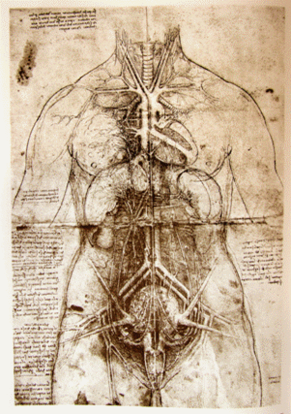
 Normal vascular anatomy. Note that the brachiocephalic
vein runs parallel and cranialto the aortic arch.
In conclusion: no dissection, no need for CT, and nothing to be worried about. Echo can be so simple.
If you want to learn more simple things about echo just visit us at 123sonography.com
Thomas Binder & the 123sonography team
Recommended articles:
Diastolic Function — A Simple Echo Approach
Assessment of Prostheses in Echocardiography
Common Mistakes #2


