Infantile or Congenital Hypertrophic Pyloric Stenosis
First published on SonoWorld
Introduction
Infantile or congenital hypertrophic pyloric stenosis is one of the most common surgical causes of vomiting in infancy. The infant may present with failure to retain feeds, persistent non-bilious vomiting after feeds, a palpable epigastric mass [which is the thickened pylorus] and dehydration [hypochloremic metabolic alkalosis due to loss of acid in the vomitus].
Pathophysiology
The circular muscle hypertrophies and this thickened muscle reduces the lumen of the pyloric channel and also elongates it. The peristaltic wave fails to pass through the narrowed pyloric channel, resulting in gastric outlet obstruction, gastric distention and possibly retrograde peristalsis.
Imaging
Ultrasound is currently the imaging modality of choice that reliably establishes the diagnosis of hypertrophic pyloric stenosis. There are various sonographic parameters that can be used to arrive at the diagnosis and include pyloric length, pyloric diameter, muscle thickness and also pyloric volume. Pyloric muscle thickness of 3 mm or greater, pyloric canal length of 17 mm or greater and the absence of the passage of a peristaltic wave through the pylorus, during the period of scanning are the diagnostic ultrasound criteria. Classically seen is:
1. A ring on transverse section, resembling a ‘doughnut’ or a ‘bulls-eye’ or a ‘target’ with the echogenic pyloric canal in the center and surrounded by the hypertrophied pyloric muscle.
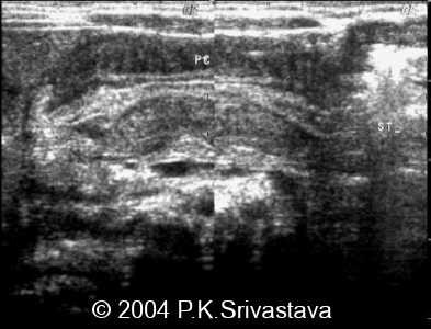
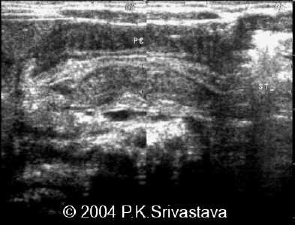
 Caption : Sagittal image of the epigastric region | Description : The pyloric channel appears significantly long and the thickened, hypoechoic muscular layers are well appreciated. The pyloric lumen is reduced and the apposed echogenic mucosal layers of both sides are noted.
2. An elongated and narrowed pyloric channel.
3. Prolapse of the redundant mucosa into the antrum creates an ‘antral nipple sign’.
4. In many cases, an elongated pylorus that lies adjacent to and just below the gall bladder provides the initial clue to the diagnosis.
5. A ‘pyloric cervix sign’ has also been demonstrated and refers to the appearance of hypertrophied pyloric musculature in longitudinal section impinging upon a fluid filled antrum.
6. The ultrasonic ‘double track sign’ is also appreciated here; however it is not specific for pyloric stenosis and can be seen in infantile pylorospasm as well.
7. Other features that may be seen are retrograde or hyper-peristaltic contractions, though none of the contractions pass through the pylorus.
The only other imaging study that may be useful for diagnosis of this condition is upper GI studies with barium. This study demonstrates the narrowed pyloric channel as a thin stream of barium passes through it [the ‘string sign’]. CT and MRI are not performed to diagnosis this condition.
Surgery [pyloromyotomy] is the treatment of choice in these patients.
1. Ito S, Tamura K, et al. Ultrasonographic diagnosis criteria using scoring for hypertrophic pyloric stenosis. J Pediatr Surg. 2000 Dec; 35(12):1714-8.
2. Levine D, Wilkes DC, Filly RA. Pylorus subjacent to the gallbladder: an additional finding in hypertrophic pyloric stenosis. J Clin Ultrasound. 1995 Sep; 23(7):425-8.
3. Reid Janet. Hypertrophic Pyloric Stenosis. http://www.emedicine.com/radio/topic358.htm#section~ultrasound
4. Hernanz-Schulman M, Dinauer P, et al. The antral nipple sign of pyloric mucosal prolapse: endoscopic correlation of a new sonographic observation in patients with pyloric stenosis. J Ultrasound Med. 1995 Apr; 14(4):283-7.
5. Cohen HL, Blumer SL, et al. The sonographic double-track sign: not pathognomonic for hypertrophic pyloric stenosis; can be seen in pylorospasm. J Ultrasound Med. 2004 May; 23(5):641-6.
6. Ball TI, Atkinson GO Jr, Gay BB Jr. Ultrasound diagnosis of hypertrophic pyloric stenosis: real-time application and the demonstration of a new sonographic sign. Radiology. 1983 May; 147(2):499-502.

