
M-Mode ultrasound imaging

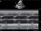
The M-mode was the preferred imaging modality in the early days of ultrasound. M-mode is defined as time motion display of the ultrasound wave along a chosen ultrasound line. It provides a monodimensional view of the heart. All of the reflectors along this line are displayed along the time axis. The advantage of the M-mode is its very high sampling rate, which results in a high time resolution so that even very rapid motions can be recorded, displayed, and measured. The disadvantage is that the ultrasound line is fixed to the tip of the ultrasound sector. It may therefore be difficult to align the M-mode perpendicular to the structures which are displayed (i.e. the septum), thus leading to false measurements.
Anatomical M-mode circumvents this limitation by reconstructing the M-mode from the 2D image (post-processing). The anatomical M-mode permits free positioning of the cursor line. However, the time resolution is significantly less than that of the conventional M-mode.
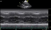
Mode tracing across the aortic sinus and the left atrium
M-mode tracing across the left ventricle
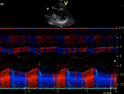
The M-mode can be combined with other imaging modalities such as color or tissue Doppler.
Curved M-mode is a color display format in which functional information (such as velocities, strain, strain rate) concerning different segments of the heart (such as the 4-chamber view) are displayed along an M-mode line which follows the myocardial walls. The M-mode line "curves" around the myocardium. Starting at the basal inferior segment it moves to the apex and back to the basal lateral wall. The functional information is color encoded.
Tissue Doppler Mode
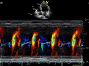
Color Doppler M-mode
The M-mode has lost much of its importance but is still valuable in certain situations.
Advantages:
- High time resolution
- Allows timing of intervals
M-mode — When is it used?
- Aorta/ left atrium (measurements, opening of aortic valve)
- Mitral/Prosthetic valve (type of valve)
- Tricuspid annular plane systolic excursion (TAPSE) for RV function
- Left/right ventricle (measurements, LV function)
- Endocarditis (motion of suspected vegetation)
- Mitral valve (Mitral stenosis)
| Other types of M-mode | Characteristics | Application |
|---|---|---|
| Anatomical M-mode | Freedom of axis | Myocardial function |
| Color Doppler M-mode | Display time motion of color | Timing of flow (i.e. flow propagation) |
| Tissue Doppler M-mode | Time motion display of functional information | Deformation and myocardial velocity analysis |
| Curved M-mode | Color-coded segmental myocardial display format that provides information about myocardial function in various segments over time | Display of myocardial velocities and deformation parameters to obtain functional information |