


Course Speakers


Curriculum
Watch free lectures
Create a free account or log in to access free lectures for this course.
No payment required.
Create free account Log inChapter 1
Starter Kit - Left ventricular function
Your entry gate to the world of echocardiography. Assessment of cardiac function made easy.
1 lesson
Lectures & Quizzes:
Chapter 2
Starter kit – Aortic stenosis
Learn how to detect the most common form of valvular heat disease. Echo is easier than using the stethoscope.
1 lesson
Lectures & Quizzes:
Chapter 3
Starter kit – Mitral regurgitation
Your first introduction to anatomy and function of the mitral valve. Learn how to use color Doppler to detect his important pathology.
1 lesson
Lectures & Quizzes:
Chapter 4
Introduction to imaging
Getting to the core of things: Instrumentation, components of an echo exam and much more…
1 lesson
Lectures & Quizzes:
Chapter 5
Imaging – parasternal views
The parasternal view – how can you find it and which views should you display.
1 lesson
Lectures & Quizzes:
Chapter 6
Imaging – apical views
The apical window- probably the most important view to the heart. A chapter full of tips and tricks.
1 lesson
Lectures & Quizzes:
Chapter 7
Imaging – subcostal views
Problems with positioning your patient? The subcostal view is a perfect alternative that allows you to also see additional structures of the heart.
1 lesson
Lectures & Quizzes:
Chapter 8
Echo in critical care conditions
This chapter will definitely change the way you manage critical care patients. Get into the driving seat of medical decision making.
2 lessons
Lectures & Quizzes:
Chapter 9
Left ventricular dysfunction
Here we dig deeper into the essentials of quantifying left ventricular function. A chapter full of practical examples.
2 lessons
Lectures & Quizzes:
Chapter 10
Acute myocardial infarction
The ECG is still first – but once you watch this chapter you will realize important echocardiography is in patients with chest pain.
1 lesson
Lectures & Quizzes:
Chapter 11
Complications of myocardial infarction
This chapter will help you save lives. You will need echo for Risk assessment and the diagnosis of myocardial rupture, ischemic VSD, right ventricular infarct and thrombi.
1 lesson
Lectures & Quizzes:
Chapter 12
Hypertrophy and myocardial disease
Hypertrophy - A frequent finding. But how do you quantify it and what are the causes? We will provide a simple roadmap.
1 lesson
Lectures & Quizzes:
Chapter 13
Fluid management
Should I give fluids or not? Echocardiography will provide the answer.
2 lessons
Lectures & Quizzes:
Chapter 14
Right heart and pulmonary hypertension
Learn how to quantify pulmonary pressure. Right heart disease is more common than you think and echo will lead you the diagnosis. From pulmonary embolism and right heart failure to tricuspid regurgitation.
1 lesson
Lectures & Quizzes:
Chapter 15
Aortic stenosis
The sequel to chapter 2, now you will also learn how to quantify aortic stenosis. We will show you how to use spectral Doppler here.
1 lesson
Lectures & Quizzes:
Chapter 16
Mitral stenosis
A common disease in many parts of the world and one of the easiest diagnosis you can make. But only if you have an echo machine.
1 lesson
Lectures & Quizzes:
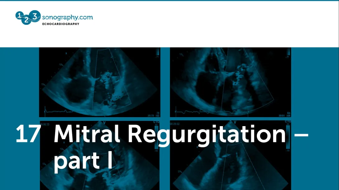
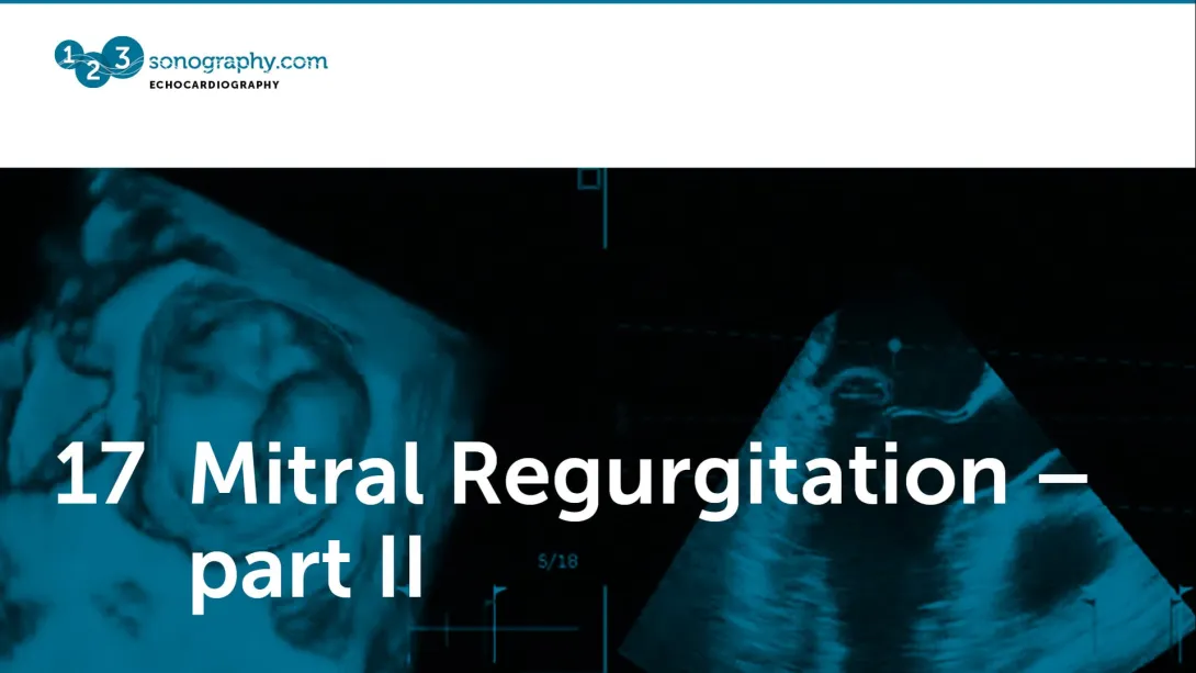
Chapter 17
Mitral regurgitation
In chapter 3 we already dealt with mitral regurgitation. Here you will see many more examples and learn why patients develop mitral regurgitation in the first place.
2 lessons
Lectures & Quizzes:
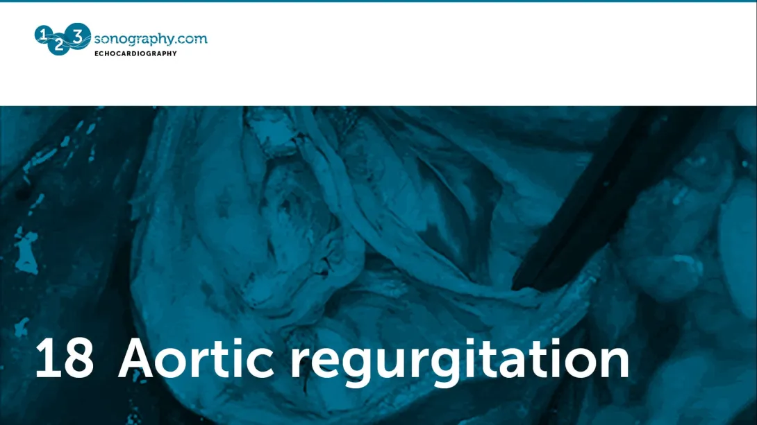
Chapter 18
Aortic regurgitation
One you know how to use color Doppler it is easy to detect aortic regurgitation. But there is more you should know about this entity if you want to manage your patients.
1 lesson
Lectures & Quizzes:
Chapter 19
Transesophageal echocardiography
Transesophageal- and transthoracic echo belong together. Learn when and how to use it. Your first step into an important modality that is not only used in cardiology.
4 lessons
Lectures & Quizzes:
Chapter 20
Heart and infection
If you don’t want to miss an endocarditis-, myocarditis or pericarditis then you will need to watch this chapter. Learn more about the incredible power of both transthoracic and transesophageal echo.
1 lesson
Lectures & Quizzes:
Chapter 21
Pericardial effusion
Detection of a pericardial effusion is one of the easiest tasks in echocardiography. This chapter will show you how to make the diagnosis and which additional information it provides.
1 lesson
Lectures & Quizzes:
Chapter 22
Aortic disease
Don’t forget to image the aorta. This lecture will explain you how and why it can save lives.
1 lesson
Lectures & Quizzes:
Chapter 23
Prosthetic valves
Over 250.000 prosthetic valves are implanted worldwide each year. So you will definitely see such patients. But how can you tell if prosthetic function is normal? We will show you in this chapter.
2 lessons
Lectures & Quizzes:
Chapter 24
Echo and cardiovascular surgery
Do you know how to assess the risk for surgery with echo? Which factors influence the postoperative outcome and what to look for in patients who underwent cardiovascular surgery? These are some of the topics discussed in this chapter.
1 lesson
Lectures & Quizzes:
Chapter 25
Premium - Methodology and Normal Findings
This chapter will provide you with in depth knowledge on what deformation is and which parameters can be measured. You will learn how Speckle Tracking works, which advantages it has to tissue Doppler and if values can be compared among different vendors. Finally, we will answer the question: Can we currently define normal values?
1 lesson
Lectures & Quizzes:
Chapter 26
Premium - How to Image Patients with ACHD
Here we teach you how to use a systemic approach when you image your patients.
2 lessons
Lectures & Quizzes:
Chapter 27
Premium - Endocarditis
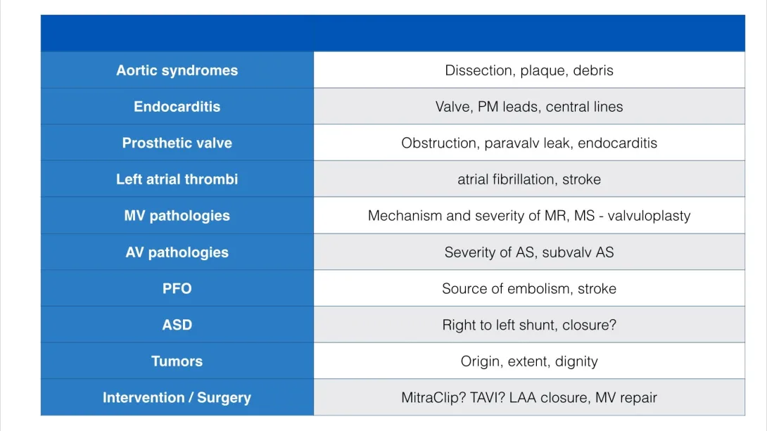
In this chapter, we cover a wide array of subjects through engaging lectures and real-world examples, including Introduction to Endocarditis, Native Valve Endocarditis, Complications of Endocarditis, Right Heart Endocarditis, Prosthetic Valve Endocarditis, Pacemaker and Central Line Endocarditis, Non-Bacterial Endocarditis, and Guiding Patient Management.
47 lessons
Lectures & Quizzes:
Chapter 28
Premium - Aortic Disease
The aorta can also be visualized with echo! In this chapter we will show you how. Diseases such as aortic aneurysms, dissection, aortic syndromes, and congenital abnormalities of the aorta will be covered.
6 lessons
Lectures & Quizzes:
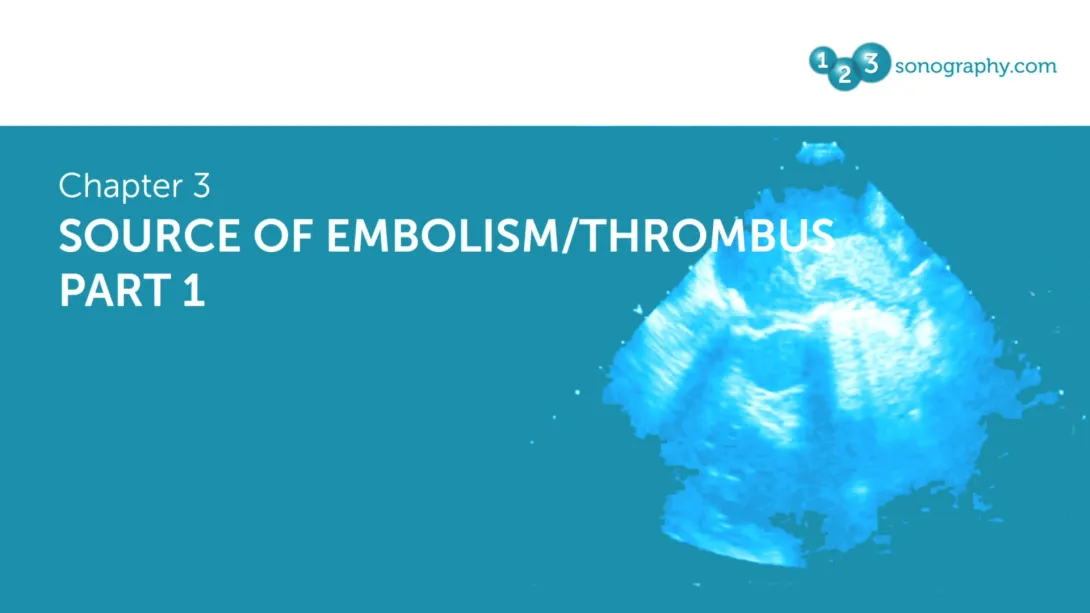
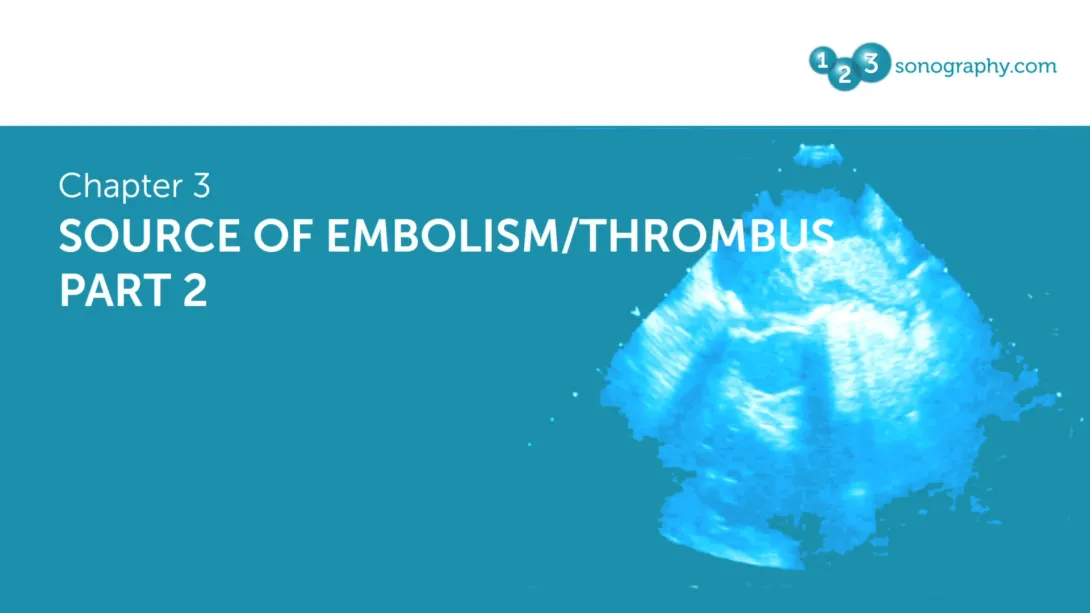
Chapter 29
Premium - Source of Embolism
Looking for a potential source of embolism is a frequent indication for a TEE.
2 lessons
Lectures & Quizzes:
Objectives

Ideal for:

Student Discount
Are you a student? Get 50% discount on this course by completing the student application form.
Get Student DiscountRecommended Blog Posts


Pricing
One-Month Access
Take the most flexible route with a monthly subscription.
You get:
- Cancellation possible anytime
- Lectures, micro- and nano-learning