
Cholelithiasis and choledocholithiasis
First published on SonoWorld
Case Presentation
35 year old female presents with right upper quadrant pain.
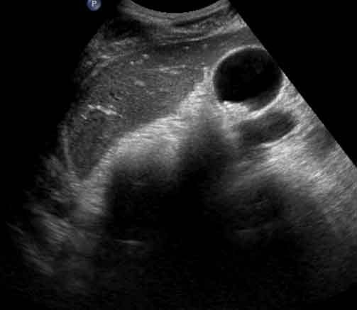 Caption: Transverse Image of Right Upper Quadrant. | Description: The gallbladder demonstrates tiny gallstones. There was mild tenderness over the gallbladder. However, there is no evidence of wall thickening or pericholecystic fluid. There was no ultrasound evidence of acute cholecystitis.
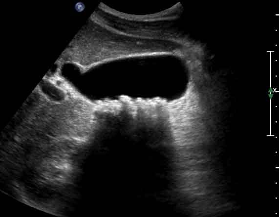 Caption: Sagittal View of Gallbladder. | Description: In this sagittal view, the gallbladder demonstrates tiny mobile gallstones with the largest measuring approximately 8 mm in diameter.
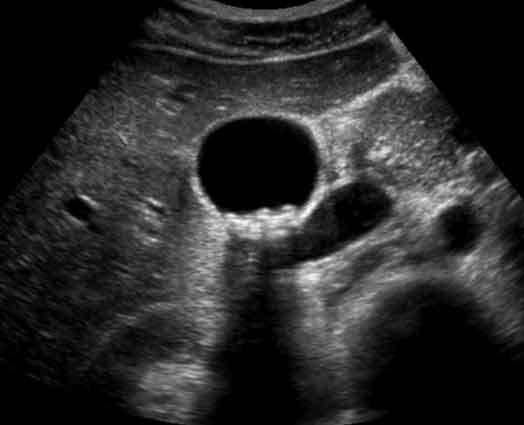 Caption: Transverse View of Gallbladder. | Description: Multiple tiny stones within the gallbladder. There is no evidence of wall thickening or pericholecystic fluid.
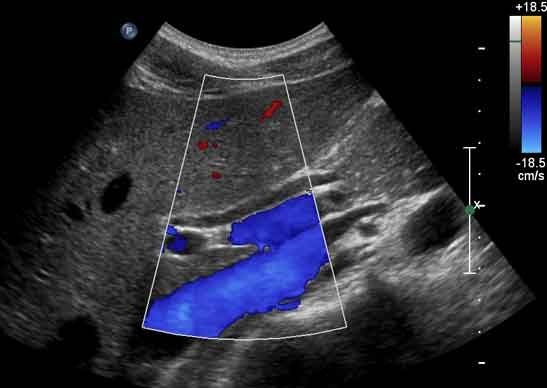 Caption: View of Common Bile Duct. | Description: The common bile duct measures 4 mm in diameter.
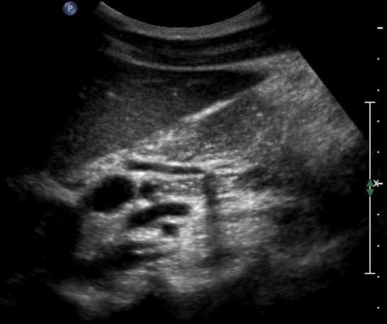 Caption: View of Common Bile Duct. | Description: View of distal portion of common bile duct shows an echogenic, shadowing stone (3 x 3mm) within the head of the pancreas.
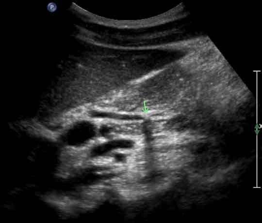 Caption: View of Common Bile Duct. | Description: An arrow indicates the position of the stone within the distal portion of the common bile duct.
Differential Diagnosis
Acute Cholecystitis.
Cholelithiasis.
Choledocholithiasis
Final Diagnosis
Cholelithiasis and choledocholithiasis