What can you find in the short axis view of the biceps tendon on the level of the intertubercular groove?
One very important structure to scan within the shoulder protocol is the long head of the biceps. Before scanning you should understand the anatomy of the long head of the biceps. So let´s recap!
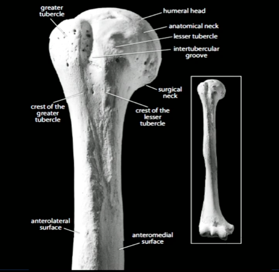
What structures can we see on the humerus?
The long head of the biceps is positioned in the intertubercular groove, which you can find in between the greater tubercle and the lesser tubercle. This groove gives the long head of the biceps its stability.
If we follow the groove along the humeral shaft to distal we see the bone gets more irregular. This is due to the insertion of the pectoralis major tendon in this region. If we follow the groove to proximal the biceps tends to be more on the medial side close to the lesser tubercle and reaches the medial humeral head to its origin the glenoid cavity.

Which muscles help to stabilize the long head of the biceps?
There are many different structures which give the long head of the biceps its stability such as the intertubercular groove and the suspensory ligament, but it is also stabilized by muscles.
The long head of the biceps lies underneath the pectoralis major muscle. This relationship creates additional stabilization. Not only the pectoralis major is involved in the stabilization of the long head of the biceps but the latissimus dorsi muscle as well. The latissimus dorsi attaches to the medial side of the dorsal humerus and lies underneath the long head of the biceps.
Together the pectoralis major and the latissimus dorsi create kind of an envelope for the long head of the biceps and stabilize it distaliy.
Now we repeated a few aspects of the anatomy, let's take a look at the sonoanatomy of the biceps!
Sonoanatomy in SAX
Case
A 54 year old man who is paraplegic after a motorcycle accident 30 years ago and has been using a wheelchair since then presents to you with pain in his left shoulder. The patient had already had surgeries on both of his shoulders. Since one year he noticed increasing pain in his left shoulder, especially during activities where he uses his left shoulder.
You perform a physical examination. There you can see the patient has no limitations in range of motion, but reports pain in the end range of external rotation on the anterior part of the left shoulder and as well during the palpation of the biceps tendon. After the complete physical exam you have the suspicion there could be something wrong with the long head of the biceps.
You perform an Ultrasound examination. What can you find in the short axis view of the biceps tendon on the level of the intertubercular groove?
A) the biceps tendon is not in the middle of the groove
B) the biceps tendon is in the middle of the groove
C) the biceps tendon tends to lie on top of the greater tubercle
D) the biceps tendon is domed and narrow
--> get the answer.


