
Category
Cases
The View from the Top

As specialist we always tend to focus on our own field of expertise and we often neglect things that we are not so familiar with. As cardiologists we tend to think that:
- patients with fever must have endocarditis,
- chest pain is caused by coronary artery disease and that
- dizziness is either related to some form of arrhythmia or aortic stenosis

So sometimes it does make sense to “broaden your view” and take a look “from the top”…
In the middle of the night
So here is what happened: A young resident paged the cardiologist on call to perform an echo on a patient who was just admitted with dyspnea. “The patient is in pulmonary edema and we need to check his left ventricular function” was the demanding call in the middle of the night. The cardiologist was not very happy to perform an exam at 3 AM. But after all - echo was essential in this setting.

When he arrived, the patient had just received his ECG which didn’t show much. All other results (chest x-ray, lab etc.) were pending. The patient had felt increasing dyspnea and was now breathing rather heavily, his oxygen saturation was 92%.
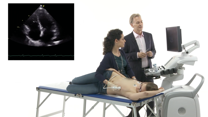
The echocardiogram
video platform video management video solutions video player Parasternal long axis view showing normal size and function of the ventricle
Searching for a pathology
Is this the echo of someone in pulmonary edema? Do you see a cause for his dyspnea? Okay, left ventricular function is normal, but maybe it is a valvular problem. But again all that color Doppler shows is trivial mitral regurgitation: This also does not explain his dyspnea.
video platform video management video solutions video player
Color Doppler showing “only” trivial mitral regurgitation
No pathology of the aortic valve was present, no pericardial effusion, a normal right ventricle and no signs of pulmonary hypertension. But why does the patient have dyspnea then? Let us take a look at the four - chamber view: Does anyone see the true problem?
video platform video management video solutions video player
Four chamber view displaying normal left ventricular function, but there is something else on the image that should catch your attention
Digging deeper We will make it easier for you and show you a four-chamber view with an even greater depth setting.
video platform video management video solutions video player
Pleural effusion
We are sure you now see what the problem is: there is a large pleural effusion lateral to the right ventricle.
video platform video management video solutions video player
Pleural imaging showing a large pleural effusion
Imaging from the back confirms the diagnosis of “bilateral” pleural effusion.

Chest x-ray confirming bilateral pleural effusion
So, the cause of dyspnea is clear. The only open question is: why does the patient have pleural effusion? As it turned out it was caused by lymphoma.
Aside from malignant causes, pleural effusions are often caused by right heart failure or infection. Less common causes include: systemic diseases (i.e., autoimmune disease) or trauma (bleeding).
This case should encourage you to look at the lungs more frequently with echo. Ultrasound is highly sensitive for the detection of even small pleural effusions. After all, why wait until you get the chest r-ray. The diagnosis can be made in a few seconds with echo - as long as you broaden your view…
If you want to broaden your knowledge of echocardiography and get “to the top”, then visit us at 123sonography.com
Recommended articles:
Genesis
Old and New
Where is Waldo (Part 1)?
Related Courses

The Echo MasterClass is a premium online training program that will bring your echo skills to…
Hours
40,3
The Echo BachelorClass provides an invaluable foundation for the practice of echocardiography…
Hours
13,1