
OB/GYN Ultrasound BachelorClass Case


A 37 year old woman comes to the emergency department at 4 am with massive cramps in her lower abdomen. Her period started two days ago. It is not unusual for her to have severe pain during her menstruation. However, this time it is even more intense and resistant to all kinds of pain medication (i.e., NSAR).
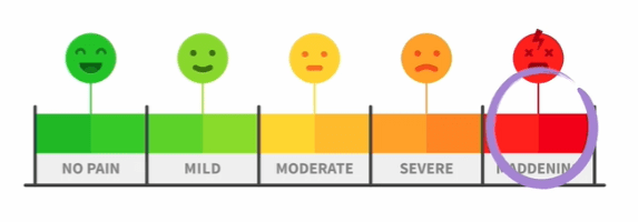
Visual analog scale (VAS) – pain rating scale
The gynecologist on duty performed a transvaginal ultrasound – Can you tell what the problem is?
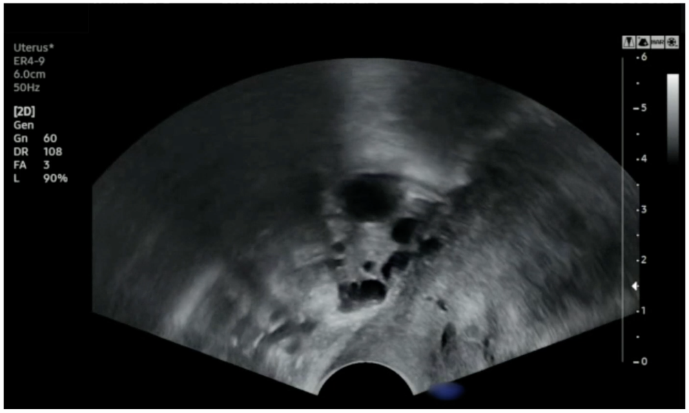
What does the ultrasound show?
You can locate an ovary (with anechoic follicles in the inner aspect) posterior to the anteverted uterus of the patient. When the gynecologist applies pressure to the probe, the ovary does not slide back and forth but stays in the center. The ovary appears to be stuck to the uterus. This is what is called a "negative sliding sign". It denotes the presence of adhesions due to inflammatory processes and is indicative of pelvic intraperitoneal endometriosis.
In contrast, a positive sliding sign is considered normal. In this case, the pelvic structures, such as the ovaries, uterus, anterior rectum, etc., displays a sliding motion toward each other when the pressure is applied to these organs with the probe.
Ultrasound saves the day!
The diagnosis is now clear based on the ultrasound examination, patient history, and severe pain (maximum score on the visual analog scale). Our patient has endometriosis. She was referred to a specialized clinic, where the suspected diagnosis was confirmed.
Which other pathologies to consider
Important differential diagnoses include pelvic inflammatory disease (PID), polycystic ovary syndrome (PCOS), and bladder infections. Uterine fibroids can also sometimes be painful.
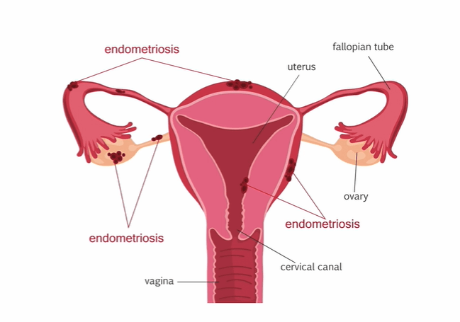
Common locations of endometriosis
We hope you enjoyed this case and if you want to learn more about our OB/GYN Ultrasound BachelorClass, make sure to register for the free lectures!
Yours,
Thomas Binder and the 123sonography team
Related Courses
This course aims to provide you with the knowledge and skills on how to do an ultrasound…

The OB/GYN Ultrasound BachelorClass is a clinical course that will provide you with in-depth…