The 4 Key "Elephants" Indicating Malignancy Risk in Thyroid Nodules: A Case of Fine Needle Aspiration
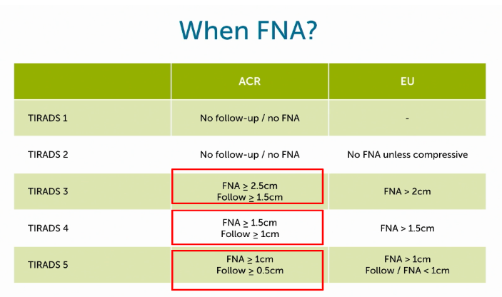
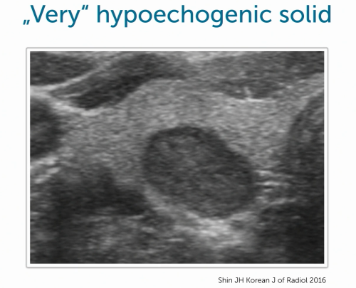
When evaluating thyroid nodules, certain "red flag" features, as categorized by the TIRADS classification, can indicate a heightened risk of malignancy. These prominent indicators should not be overlooked:
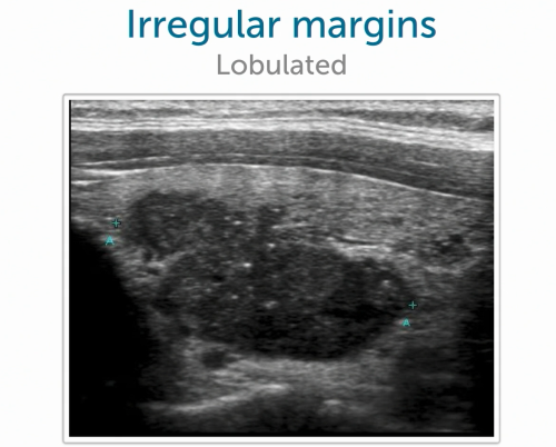
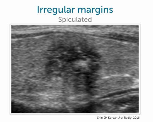
3. Irregular Borders and Shape
Lobulated or spiculated (spiky) borders are signs of malignancy. Irregular, poorly defined edges suggest tumor growth extending into surrounding thyroid tissue. It is essential to assess these nodules
three-dimensionally to capture such a spiculated or lobulated shape. Here are two examples:
4. "Taller-than-Wide" Shape
Nodules that grow more in-depth than in width, giving an "egg-like" shape, raise suspicion of malignancy. Nodules extending towards the posterior thyroid regions are particularly concerning. Refer to Image 4 to visualize this characteristic.
While these features are significant indicators, assessing malignancy risk requires a comprehensive approach, as covered in detail in our extensive Thyroid Ultrasound MasterClass. Start the course now with our free lectures!
Case Presentation: Fine Needle Aspiration of a Thyroid Nodule
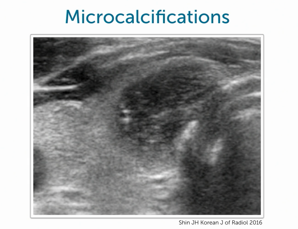
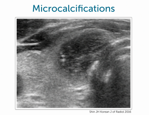
In the following case, we applied these malignancy criteria in assessing the patient's nodule. The patient was referred for a thyroid ultrasound after noticing a 4 cm lump in her neck. Ultrasound revealed a cystic structure with a solid component measuring approximately 2 cm, displaying an irregular, inhomogeneous texture and the presence of microcalcifications. Additionally, the patient reported discomfort due to tissue compression from the nodule.
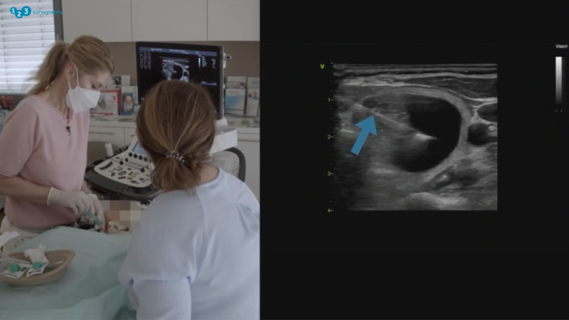
The patient was referred for a thyroid ultrasound after noticing a 4 cm lump in her neck. Ultrasound revealed a cystic structure with a solid component measuring approximately 2 cm, displaying an irregular, inhomogeneous texture and the presence of microcalcifications. Additionally, the patient reported discomfort due to tissue compression from the nodule.
Procedure Details
Performing FNA was challenging due to the mixed cystic-solid nature of the nodule, often requiring a two-step approach to address both components. During the procedure, we were able to extract some of the cyst fluid. However, due to its consistency, residual material remained, which may lead to future refilling.
Follow-Up and Potential Outcomes
Should pathology confirm a papillary carcinoma, the patient will be referred for surgery. If the nodule is benign yet symptomatic due to recurring fluid accumulation, surgical intervention might still be considered to relieve persistent discomfort.
Final Considerations
FNA is a semi-invasive procedure, and patients can experience anxiety before, during, and even some pain afterwards. It is essential to communicate clearly with patients to help manage expectations and reduce procedural anxiety. The entire process of Fine Needle Aspiration (FNA) is crucial, from informing the patient about the necessity of the procedure to their departure after the process and even concerning the biopsy results.


