
How to diagnose pneumonia in yourself


Ben Thal, a main speaker in our upcoming course "Prehospital Point-of-Care Ultrasound", shares a case where his colleague Chiara unexpectedly became a super exciting pPOCUS case.
Chiara, a medical student, unexpectedly diagnosed herself with pneumonia using a bedside ultrasound device from her e-learning project with us. Initially suspecting acute tonsillitis due to severe sore throat and other symptoms, her condition didn't improve with standard medication. Using point-of-care ultrasound (pPOCUS), she discovered signs of pneumonia, underscoring the tool's value in medical diagnosis and education.
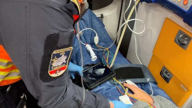
Admittedly, bedside ultrasound for medical students doesn’t necessarily mean having an ultrasound device next to the bed at all times. But for Chiara, this was the case, partly because we use every free minute to collect scans for our e-learning project with 123sonography. For this, we have two portable devices in use. I have one of them and carry it with me for almost all of my medical engagements (e.g., during senior physician duties at the Vienna Professional Rescue Service, in my practices, on the helicopter…) and the other is with her, mainly used in her life as a medical student, such as in her job as a sonography tutor, in the practices where she works, or during my emergency medical services where she accompanies me. Thus, one evening, the device was once again lying on the charging station in her apartment, and one fine evening in December, she unexpectedly became a super exciting pPOCUS case herself.
However, the story began two days earlier when I suddenly received a message from Chiara saying she had a sore throat and her self-analysis as a medical student suggested it could only be acute tonsillitis. She might have overdone it a bit in sports in the previous days (and when an ex-athlete tells you that, you can imagine that the ambition bird had a good day). Anyway, her throat looked awful: swollen, coated tonsils, swollen lymph nodes, petechiae on the soft palate, fever up to 39°C, no cough. Overall, a Centor Score of 4 and, for me, it was obvious to agree unreservedly to her request for a pharmacological frontal attack and to send the appropriate prescription. So, I sent her a nice Advent prescription via WhatsApp, including Ibuprofen, single-shot cortisone, and a broad-spectrum penicillin, generously dosed, especially considering the upcoming holidays. Streptococci, take cover.
Most patients are actually very well served with this cocktail. Apparently not Chiara!
Two days later, an evening call. “Hey, I’ve suddenly developed a cough since noon and pain right parasternal in the area of the 2nd intercostal space. I thought I’d hold the ultrasound probe there, and it looks super weird. Can you take a look?” She then sends me the first loops that speak for themselves.
 As more fluid accumulates in the lung, sections of the lung may become entirely filled with fluid, resulting in consolidation. This phenomenon is frequently observed in cases of pneumonia. It’s important to note that consolidation can also result from atelectasis due to airway obstruction (such as a mucous plug) or external compression (such as a large pleural effusion).
What we see here is a small, pleural consolidation, most likely in the sense of infiltration. Since this ultrasound finding could theoretically also be a peripheral pulmonary embolism, additional images with the color Doppler are supplied. These, however, show no abnormalities, and the slender right ventricle speaks at least against a large pulmonary embolism.
 A slender right ventricle supports the suspicion that at least it is not a large pulmonary embolism (but does not rule it out).
Moreover, descending lobular pneumonia, according to literature, is most often caused by pneumococci and/or other streptococci (source: Amboss.com). Due to her athletic habitus, the overall good general condition, and a patient slightly refusing hospitalization, we decide to continue therapy at home with a ban on sports, bed rest, increasing the antibiotic therapy, and some other supportive measures. When I visited her two days later, she was already feeling much better. Scanning her again, the findings still look relatively unchanged. But watch how the B-pattern remains almost exclusively limited to the 2nd and 3rd segments of the right upper lobe and immediately transitions into an A-pattern further caudally.
 As the linear transducer was slid vertically down from the right parasternal 2nd intercostal space (ICR) the B-lines spontaneously transitioned into an A-pattern.
What can we learn from this case in the spirit of our pPOCUS mission?
- Chiara had never scanned pneumonia before – and yet hit the bull’s eye immediately. Conclusion: Take the ultrasound probe, hold it, read up, have it verified by experienced colleagues if necessary!
- Incipient lobular pneumonias can occur following streptococcal angina
- B-lines can be limited to small regions in pneumonias, e.g., in the marginal areas of the consolidation
And Chiara is happy that, apart from the cool story, there are no residues to complain about.
Do you want to learn how to perform prehospital point-of-care ultrasound yourself?
We have already published a few chapters of our course with more to come. Get yourself a 30% discount now and Lifetime Access to the course – until July 31st, 2024, only!
Related Courses
This course will equip you with expertise in applying POCUS across diverse prehospital scenarios…