Electrosonography
Just the other day, we were dwelling over an echo trying to figure out if the patient had an ASD or not. We just could not tell from the images. Do you know what ultimately helped us? A simple ECG. The fact that the ECG was completely normal and that not even an incomplete right bundle branch was present, practically rules out an atrial septal defect.
The ECG is quite helpful in many situations. I remember a colleague who always looked at the ECG first before he came to the decision whether regional wall motion abnormalities were present. Some might think it is a way of “cheating“ or that it biases your echo interpretation. We think it is simply a smart way of coming to the right conclusion.
So today I would like to test YOUR ECG skills. Don‘t worry, it will also have to do with echocardiography. I will present you with four ECGs and four echo loops. Lets see if you can match the ECG to the echocardiogram of the same patient.
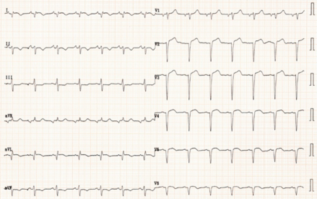
Here are the electrocardiograms:


And these are the echocardiograms (click images to see videos).
1
video platform video management video solutions video player
2
video platform video management video solutions video player
3
video platform video management video solutions video player
4
video platform video management video solutions video player
Take your time and try to come up with the correct echo diagnosis first. Then look at the key findings in the ECG.
Here are the correct answers:

ECG A matches echo 1: The patient has obstructive hypertrophic cardiomyopathy (HOCMP). In such patients you will typically find signs of left ventricular hypertrophy and negative T waves in the precordial leads.
ECG B matches echo 2: The patient has a large left ventricular aneurysm following anterior myocardial infarction.The ECG therefore shows persistent ST-elevations.
ECG C matches echo 3: The correct diagnosis here is severe pulmonary hypertension with a dilated right ventricle and reduced right ventricular function. A very common ECG finding is that of a right ventricular strain pattern and signs of right ventricular hypertrophy.
ECG D matches echo 4: What you see here is a short axis view displaying the right ventricular outflow tract. Note that it is dilated and aneurysmatic, which suggests right arrhythmogenic right ventricular dysplasia (ARVD). Such patients are at risk for arrhythmias such as ventricular tachycardia.
These are only a few examples of how the ECG can provide clues to the correct diagnosis. Remember, the echocardiogram is only one piece of the puzzle. And the more pieces you have the easier it will be to see the entire picture.
To understand more on how to make the right diagnosis visit us at 123sonography.com
Recommended articles:
Mausi - Hypertrophic cardiomyopathy
Echo Imaging of the ASD
function advagg_mod_1() { // Count how many times this function is called. advagg_mod_1.count = ++advagg_mod_1.count || 1; try { if (advagg_mod_1.count <= 40) { // Set this to 100 so that this function only runs once. advagg_mod_1.count = 100; } } catch(e) { if (advagg_mod_1.count >= 40) { // Throw the exception if this still fails after running 40 times. throw e; } else { // Try again in 250 ms. window.setTimeout(advagg_mod_1, 250); } } } function advagg_mod_1_check() { if (window.jQuery && window.Drupal && window.Drupal.settings) { advagg_mod_1(); } else { window.setTimeout(advagg_mod_1_check, 250); } } advagg_mod_1_check();

