Detecting the Missing Link
-->
We all know Sherlock Holmes or have watched inspector Columbo solve complex crimes and puzzles. We have admired their ingenuity, their ability to see the invisible. When we watched Sherlock Holmes movies or read the books, we realized that he observed details that went unnoticed by his constant companion - Dr. Watson. Holmes knew exactly what seemingly unimportant details needed his attention. Very often, we as “diagnosticians” are in a similar situation. We need to evaluate a cardiac structure, pathology or physiology but the essential information is missing due to poor image quality, technical difficulties or similar. In this situation, we need to learn about the seemingly unimportant features that will solve the mystery; we need to leave Dr. Watson behind and become Sherlock Holmes. In this post, we want to provide a few clues that will help you to reach echo inspector stardome....
What if you want to assess left ventricular function (LVF) but the endocardium is not visible.
There are a couple of things you should pay attention to....
- Sometimes, the entire endocardium is not visible in the standard views - switch to atypical views and optimize for individual segement.
- If this approach does not work, use LV contrast agents.
- Also look at diastolic function - if it is normal, LVF also has to be normal. Patients with LV systolic dysfunction always have some degree of diastolic dysfunction.
What if you want to assess regional wall motion abnormalities but image quality is poor.
- Look at the shape of the ventricle. Very often, the ventricle becomes distorted after myocardial infarction, even in patients that don’t have an aneurysm.
- Furthermore, you should have a look at the patient's ECG. Does it show any Q-waves, slow R-progression, ST-segment abnormalities or negative T-waves? We are big fans of using all the clinical information available for coming up with a well informed echo report and you should be too.
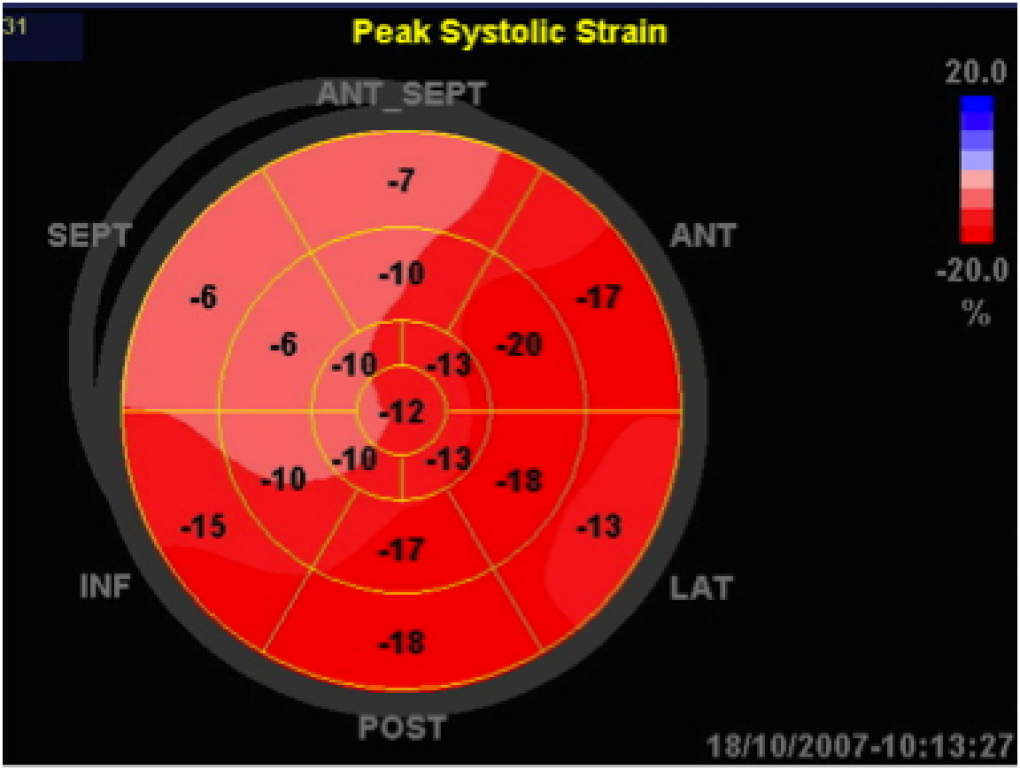
- If you are comfortable with newer echo techniques, we advise you to also use speckle tracking. We use it on almost all of our patients and it works like a charm. Most machines will even provide you with a bull's eye map very similar to SPECT (i.e., scintigraphy) reports. See an example of a bull's eye of an anterior wall infarction below.

You need to know if the patient has pulmonary hypertension but there is no adequate tricuspid regurgitation signal.
- One of the most indicative signs of pulmonary hypertension in this situation is the presence of a D-shaped left ventricle in the parasternal short axis view . A D-shaped left ventricle in the abscence of severe tricuspid regurgitation is highly indicative of pulmonary hypertension.
video platform video management video solutions video player
- Alternatively, you can also measure the pulmonary velocity acceleration time or PVAT. If it is below 110 ms, the pulmonary pressure is possibly elevated. If it is below 80 ms, you can be almost certain that pulmonary hypertension is present. The PVAT is the time interval between the beginning of the outflow signal through the right ventricular outflow tract and its peak (see image). Also look for a notch in the downslope of the curve, this also indicated pulmonary hypertension.
 This is it for now. Watch out for more investigative advice to come. We hope you enjoyed this newsletter. Let us know if you have any topic requests for future newsletters.
 Take good care,
your 123sonography Team
Recommended articles:
Electrosonography
Optimized 4-Chamber View: the Right Ventricle

