
Common Mistakes #2

We recently sent out a newsletter on common mistakes in echocardiography where we explained how to adjust your machine settings in order to optimize your image. This time we would like to focus on other "practical issues" of echocardiography that will help you to get the maximum benefit for your clinical practice.

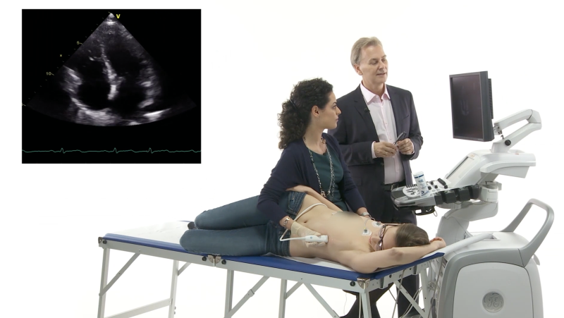
Positioning the patient:
Examining the patient in the correct position is very important. But it is often neglected. Here are a few tips:
- For the parasternal window you need to really position the patient in a left lateral position; to image the pulmonary artery move the patient even further to the left.
- For the apical window, the patient should still be positioned in a left lateral position but not as much as for the parasternal window. Simply because you cannot get to the apex if the patient is too far on his left.
- To get a good suprasternal view, let the patient’s head “hang over the bed“ and have the patient extend his head back as far a possible. Image during expiration!
- For the subcostal position, the patient should be lying on his back. To relax the abdominal muscles, let the patient bend his knees.
Respiration Friend and Foe?
Even though respiration makes imaging more difficult, you can use it to your own advantage!
- Image quality can actually improve with inspiration! This is especially true when visualizing the inferior wall in the 2 chamber view.
- Move the transducer with the movements of the chest during respiration!
- For most other views, let the patient exhale. Sometimes, letting the patient hold their breath (either in inspiration or in expiration) also helps.
- Let the patient slowly inhale while you move the transducer towards the xiphoid when you are performing a subcostal view.
- Talking to the patient is important but ask the patient NOT to talk during the exam!
- One last point: don’t stigmatize the patient with how he/she should not breath too much. The chances are that patients will breath more consciously and therefore deeper (making imaging all together more difficult).
Optimize the 2D Image
before you use other modalities like MMode, color, spectral- or tissue Doppler. Poor 2D image quality also means poor Doppler and MMode quality! If you lose the signal go back to the 2D image. Keep in mind: 2D always comes first!
Use a systematic approach!
Novice investigators tend to jump around between different views and windows. Experts on the other hand use a systematic approach. Start with the parasternal window then proceed to the apical window and end with the subcostal window. Perform all the different modalities (Doppler, MMode etc) that can be obtained in one window before you go to the next window. That way you won’t forget important parts of the exam. But, go back and obtain additional information if things are unclear. This will also shorten the exam time. And last but not least: be brave! Experiment and use atypical views! This often allows you to optimize your image to the region that you want to interpret .
In the next newsletter we will discuss documentation. Also a very important issue not only for the patient but also for legal reasons.
your 123sonography team
Recommended articles:
Echo Genius - Writing the Report (Part 1)
Echo Genius - Writing the Report (Part 2)
Echo Genius - Image Documentation
Improving Your Scanning Skills
7 golden rules of echocardiography
Free Content
Our fast and efficient learning techniques help you practice the nuts and bolts of ultrasound. Start now - it's free!Related Courses
The Echo BachelorClass provides an invaluable foundation for the practice of echocardiography…