
Beyond the Basics: Vascular Lower Extremity BachelorClass

Patient case
Mastering lower extremity duplex ultrasound is a crucial cornerstone in modern ultrasound diagnostics, offering a swift and non-invasive way to assess vascular health. Beyond its clinical utility, this skill holds profound implications from a global health perspective, particularly when it comes to deep vein thrombosis (DVT) and its "bigger sibling" pulmonary embolism (PE). Lower extremity duplex ultrasound enables clinicians to visually inspect the deep veins of the legs, detecting the presence of blood clots with remarkable accuracy and in real-time. This means that patients presenting with symptoms suggestive of DVT can undergo immediate evaluation, leading to timely interventions that could alleviate or even avert the devastating consequences of PE.
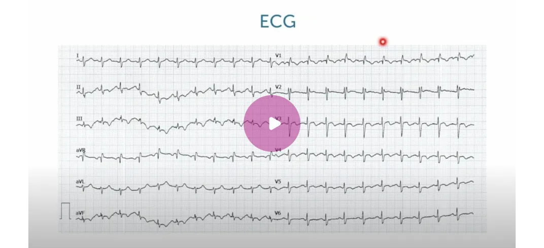
Today, we’d like to present a case of a 23 year-old woman who had just returned from a long plane trip and experienced syncope shortly after her return. Her electrocardiogram (ECG) showed an incomplete right bundle branch block and T-wave abnormalities.
Her echo study revealed a dilated right ventricle and flattening of the interventricular septum, raising suspicions of pulmonary embolism (PE), especially when considering the recent long-distance journey. The subsequent CT study confirmed the suspicion depicting a massive, bilateral pulmonary embolism, and the patient was put on systemic thrombolysis.
Did you realize how valuable an echo exam can be to corroborate your suspicion of a PE?
So now we have a confirmed diagnosis, but where did the embolus initially come from?
Beyond 3-point Compression Sonography – What about the sub-popliteal level?
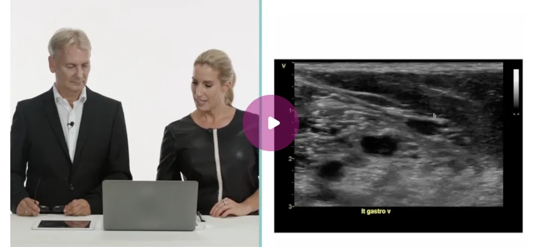
Interestingly, the initial emergency duplex ultrasound evaluation in the emergency department (three-point compression sonography) did not reveal evidence of blood clots down until the popliteal level. Upon reevaluation a few days later, a dilated, non-compressible gastrocnemius vein with extensive thrombus formation at the confluence of the popliteal vein was identified!
To summarize, we know that the pulmonary embolism was either caused by a large thrombus formation in a larger deep vein that was not visible on ultrasound because it had already traveled to the lungs, or it could actually have been caused by the extensive blood clot formation in the gastrocnemius vein detected on the second, more detailed duplex study.
First, this case underscores the significance of mastering lower extremity duplex ultrasound beyond three-point compression sonography. In some situations, the entire deep venous system, i.e., the common femoral veins, the popliteal vein, as well as the peroneal, anterior tibial, posterior tibial, soleus, and gastrocnemius veins, has to be assessed.
Did you know that we just started our Ultrasound Happy Hours with the Vascular Lower Extremity BachelorClass? Just like in this case, learning how to scan the lower extremities enables you to help diagnose life-threatening conditions like DVT and many other vascular pathologies accurately. Therefore, we are currently offering a 50% discount on the Vascular Lower Extremity BachelorClass, which will teach you just that and will ultimately improve your patient’s outcomes and well-being! Get your access now!
Related Courses

With the Vascular Lower Extremity BachelorClass, you will gain a strong foundation and…