
Can YOU do it?
Doing something for the first time can be a lot of fun. Going to your first party, traveling to a place you have never been before, or the first time you date someone. However, your first resuscitation, first intubation, or seeing your first critically ill patient can be a stressful and rather unpleasant experience. You are putting your patient and your reputation at risk. How can you be prepared for such a situation? Do you really wish to practice on your patients? Not a good idea. The solution is simple: use simulators to learn. Pilots use flight simulators to learn how to fly a plane and cope with critical situations. If it can be done in aeronautics, why not in medicine?
It can be done
Simulators are already being applied in many areas of medicine. One can practice medical procedures on phantoms, surgery on animal specimens, or even use ultrasound simulators to learn how to perform sonographic investigations. Recently I had the opportunity to visit the Palmetto Health Simulation Center in Columbia, South Carolina. Eric Brown and his team have created a fantastic facility, full of modern technology, to teach a clinician how to deal with a wide variety of emergencies.
 Thomas Cook and Eric Brown at the Palmetto Health
Simulation Center.
What an experience it was to see young students and residents use this unique opportunity to learn - with absolutely no risk of harming patients. In the following I present one of the scenarios that were simulated. Let's see if you can master the following case:
Syncope and shortness of breath
Here is the information that was given to the team:An elderly woman presents with her husband at the emergency department after she passed out in a nearby mall. Her husband caught her in his arms and she quickly regained consciousness. She was able to walk to the car with some difficulty and noted increasing difficulty with breathing. She had a history of active breast cancer and lumpectomy, and is taking metformin, omeprazole and naproxen.
123 action
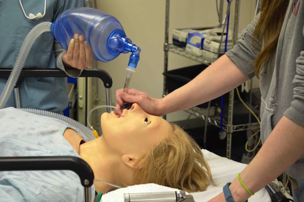
Let us now watch the scenario created and presented by Thomas Cook, based on a real patient. Professional narrators, actors, and a manikin assist in making the scenario as realistic as possible.
Then they are on their own…
So far so good
Here is our summary: our patient is critically ill, gasping for air (her respiratory rate is 35/min), her heart rate is 161/min, she is severely hypotensive (90/60 mmHg), and her oxygen saturation is only 78% (with oxygen = 86%). One leg is swollen.

The monitor shortly after the patient received oxygen.
The team provides oxygen and places two large-bore IV´s. What comes to your mind? Do you sense the problem? What test would you perform first? Some of you might advocate an ECG, a chest X-ray, or a lab test. The correct test is ultrasound, specifically echocardiography. This is exactly what the team suggests. Here it is:
Echocardiography solves the case.
I would say the echo is diagnostic. The patient most likely has acute pulmonary embolism. The echo shows severe right ventricular enlargement and poor right ventricular function. Her inferior vena cava (IVC) is severely enlarged with no changes on respiration. This is a clear case of severe acute pressure overload. Why can it not be chronic pressure overload (severe pulmonary hypertension) with sudden right heart failure? Simple: she has no right ventricular hypertrophy. To highlight the importance of echo: within 2 minutes you have the diagnosis. The high pre-test probability (symptoms, oxygen saturation, a history of malignancy, swollen leg etc.) together with the typical echo findings are diagnostic. The next step is heparin and TPA. Your only hope lies in dissolving the thrombus as quickly as possible.
Why don’t you give inotropic drugs? Because she does not have a “left heart problem”: look at her echo. Fluids are also not suitable. Her IVC is wide and hypotension is simply caused by the fact that clots are clogging her circulation. As a matter of fact, fluids will enlarge the right ventricle further. This will compress the left ventricle and reduce cardiac output. Now that she is intubated and her blood pressure is stabilized with vasopressors, let's look at the other findings:
 ECG showing sinus tachycardia and a typical
right heart strain pattern.
The ECG is very typical for acute pulmonary embolism. In fact, it shows all the typical features one would expect. Tachycardia, right axis deviation, right bundle branch block, and a right ventricular strain pattern.

The chest X-ray shows a prominent PA, no effusion
and no pulmonary congestion.
The diagnosis is confirmed with pulmonary CT, which shows massive emboli in both pulmonary arteries:

Saddle pulmonary embolus (bilateral pulmonary embolism).
The team and the ultrasound investigation saved the patient. How did the patient do in real life? She survived. And why? Because the team in charge was well trained - with the help of simulators among other aids.
If you think your friends should know how important ultrasound is and if you find our method of teaching stimulating, just forward this teaching mail to others.
Best
Thomas PS: Do you want to read more on pulmonary embolism / pulmonary hypertension and right heart disease? Here are a few Blog entries that you might want to watch:
Three stories one plot
Echocardiographic finding of pulmonary hypertension
Precapillary pulmonary hypertension
PPS: Please leave you comments below we want to know what YOU think.