
1-2-3 - How many do you see?
Have you ever been so drunk after a party that you lost all your elementary (and human) abilities like standing upright, talking sensibly, or counting properly? On those very occasions, however, you gain new or partly new abilities like crawling, prattling, or laughing uncontrollably while crying! Isn't that amazing? You know, echo sometimes gives you the impression of being drunk. You see things which are not there, shouldn’t be there, or should simply look different. Go through the following echo case and you'll get what I mean.
Our case
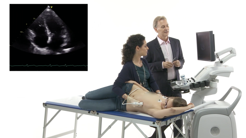
Sandy is a 19-year-old teenager who has been dealing with physicians since the day she was born. Why? Simple: her parents are doctors - cardiologists to be precise! The day Sandy’s mother received her new echo scanner, the girl ended up on a medical bed in her mother’s private office, scheduled to undergo an echo investigation. It was supposed to be a normal heart. Sandy was young and had no symptoms whatever. However, one thing in her heart looked too big. Curious? Take a look yourself.
TTE, parasternal long-axis view. The ascending aorta
(Aa) looks enlarged.
Sandy’s ascending aorta was 39 mm and looked too large for her body surface area (BSA, 1.8 mq; the index for the ascending aorta is 21.6 mm/mq). Concerned by these findings and eager to discover the cause, Sandy’s parents sent her to our lab for a second opinion.
Our finding
In the meantime, the results of the screening tests for connective tissue disease had arrived - they were negative. Apart from confirming her enlarged ascending aorta, we looked at the aortic valve. After all, a dilated aorta is often seen in the presence of a bicuspid valve. Now let’s see if you don’t feel like you were drunk when looking at this loop. How many cusps do you see in Sandy’s aortic valve?
TTE, parasternal short-axis view. Aortic cusps.
Three, right? The question is: do all cusps separate during systole? We went through the image loop frame by frame, and this ensured me that the cusps did. But then why is the aorta dilated? We discussed this with her mother, the referring physician, and finally decided to perform a TEE study.
Let’s test your ability
Here is a TEE still frame of Sandy’s aortic valve. What is your opinion now? Tricuspid or not?

TEE, short-axis view focusing on the aortic valve.
If you answered the question with a “yes”, please lie down on the ground and do 10 pushups as a penalty… you are wrong! If your answer was “no” please be ashamed as well, and do 10 pushups…because your answer is wrong! If you answered, “I cannot establish the diagnosis from a still image," well congratulations, you are right! You always need to assess the aortic valve in motion. You need to observe how it opens and closes. This is best done with multiple echo views.
Our analysis on Sandy’s TEE
Let’s have a look at the following loop:
TEE, short-axis view focusing on the aortic valve.
The same valve. Does TEE help you solve the case now? On this short-axis view you see that the aortic cusps separate from each other during systole. However, not entirely - there is a very small region where the cusps are fused. The fused portion of the valve is also known as the raphe. Look at the shape of the aortic orifice during systole. Does the latter resemble a triangle (tricuspid valve) or is it more elliptical (bicuspid valve)? I think it is somewhere in-between. This is not the case, however… look at the sinuses of Valsalva. Sometimes they differ from each other in bicuspid aortic valves. Here the difference isn't very great, actually.
In conclusion… were we drunk during the TTE?
No, we weren’t. Detecting bicuspid valves can be quite difficult at times, especially in valves which are not truly bicuspid but only “functionally bicuspid”. Where three cusps are present but two are completely or partially fused (as in Sandy's valve), you may need a TEE to make the correct diagnosis.
Our teaching points
What are the tips and tricks on how to detect congenital abnormal valves? First, look carefully at the opening and closing motion of the valve. Second, look at the shape of the valve during systole. Third, look for doming of the valve (on a long-axis view) and finally, be suspicious. In the setting of aortic root dilatation, it cannot be explained otherwise. Remember: no matter how severe the congenital malformation of the aortic valve is, whether it is stenotic or not, functional bicuspid or truly bicuspid, unicuspid or quadriscupid, patients are all at risk of developing aortic stenosis and aortic aneurysm. Thus, it is by no means trivial to know whether Sandy's valve has a congenital abnormality or not. Now that we knew, we told Sandy to undergo a follow-up study in a few years to see whether her aortic root dilatation had progressed and to monitor the gradients across her aortic valve.
By the way, keep learning from our courses on 123sonography. Sober or not, we'll make sure you detect all there is to see.

Best,
Anna
Related Courses
The Echo MasterClass is a premium online training program that will bring your echo skills to…
The Echo BachelorClass provides an invaluable foundation for the practice of echocardiography…