
1.8.1.1 Pulsed Wave Doppler (PW-Doppler)

Pulsed wave Doppler employs elements of the transducer that send as well as receive signals. Ultrasound is emitted in "pulses" between these pulses. The same transducer element receives the reflected signal. As every emitted pulse is paired with a corresponding return signal, it is possible to determine where the reflection has occurred and calculate the distance of the "reflector". In reverse, by defining a distinct region of interest (sample volume) one is able to display returning signals from specific regions in the heart. Thus, PW Doppler has the advantage of being "site specific" as regards Doppler information. However, PW Doppler has a major drawback: it cannot correctly depict higher velocities (usually above 1.5 - 1.7m/sec). To understand the reasons we have to first address aliasing phenomena.
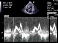
Pulsed Wave Doppler Tracing of mitral inflow
Aliasing phenomena
Aliasing is best explained by the analogy of a clock. Let us assume you are observing a clock at 15-minute intervals. Obviously you will note that the hands move in clockwise direction. However, if you were to use a longer observation interval of 40 minutes you would gain the impression that the hands move in counter-clockwise direction. This is exactly what happens with pulsed-wave Doppler when the velocity is too high. The interval of the ultrasound pulses (pulse repetition interval) is the time interval between your observations of the clock. When the pulse repetition interval is too long relative to the velocity of blood flow (in other words, when the hands of the clock move a long distance between observation periods), it will not be possible to determine the direction of blood flow.
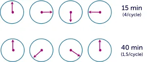
Clock analogy explaining the aliasing phenomenon
Instead of using the "pulse interval" the scanner displays pulse repetition frequency (PRF), which is the number of pulses within one second. The PRF limit within which aliasing occurs is known as the aliasing or the Nyquist limit.
Specifically, aliasing occurs when the velocity is more than one half of the pulse repetition frequency. In this case velocities above this limit will be displayed on the tracing opposite to the true direction of blood flow.
As mentioned earlier, PRF is an important aspect. To a certain degree the PRF can be increased to permit higher velocities to be displayed. However, the maximal PRF depends on imaging depth (the depth position of the sample volume). The higher the depth of the sample volume, the longer the PRF must be (as the ultrasound wave takes longer to travel, one needs longer intervals to observe the returning of the wave). Therefore, the maximal velocity which can be displayed with pulse-wave Doppler decreases as the sample volume is positioned farther away from the transducer. Other factors that influence aliasing are:
- Depth
- Width of sample volume
- Velocity
- Doppler frequency
Aliasing will occur when the velocity exceeds the Nyquist limit. The Nyquist limit is equal to one half of the pulse repetition frequency. Use the baseline shift to "stretch" the Nyquist limit.
In summary, the advantage of PW Doppler is that it is site specific. Its disadvantage is that higher velocities cannot be measured.