12.6 Jet direction and mechanism of MR
The direction of the jet is an important indicator of the mechanism of mitral regurgitation. You will encounter situations in which the pathology itself is not visible. Your interpretation will be based on the direction of the jet alone. This section tells you how the mechanism influences the direction of the jet and the views in which the jet is seen best.
Bi-leaflet mitral valve prolapse
The symmetry of the valve is preserved in bi-leaflet prolapse. Thus, the direction of the jet is quite central. Deviations may occur, depending on the degree of prolapse (anterior vs. posterior).Thus the jet may be directed more laterally or medially. Use a four-chamber view to visualize the direction of the jet.
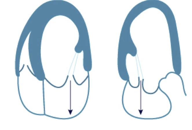

Prolapse or flail posterior leaflet
Here the jet is directed anteriorly and medially towards the interventricular septum. The jet may be very eccentric and therefore difficult to see. Use a four- or three-chamber view as well as atypical views to visualize the direction of the jet.



Prolapse of the posterior leaflet on a coronary sinus view
Prolapse of the posterior leaflet on color Doppler
 
Flail of the posterior leaflet with and without color Eccentric jets may impinge on the wall and encircle the left atrium.Prolapse or flail anterior leaflet
The jet is directed posteriorly and laterally here. This is best seen on a four- or three-chamber view. Additionally use a parasternal long-axis view to demonstrate very eccentric posterior jets.

When an entire leaflet (base to tip) is flail, the leaflet may swing open completely (similar to a saloon door). In this case the direction of the jet will follow the angle of the flail leaflet; it may be directed more laterally or even along the lateral left atrial wall.

Saloon door effect: The posterior leaflet is "completely" flail
As the posterior leaflet swings open completely during diastole, the jet is directed more laterally.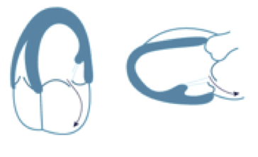

 
Flail anterior leaflet with and without colorPerforated leaflets
Leaflet perforation usually occurs as a result of endocarditis. The anterior leaflet is more frequently involved. Typically the origin of the jet is located more towards the base of the leaflet. The jet appears to pass straight through the valve. Use a parasternal long-axis, three- or four-chamber view to identify the pathology (with and without color Doppler).
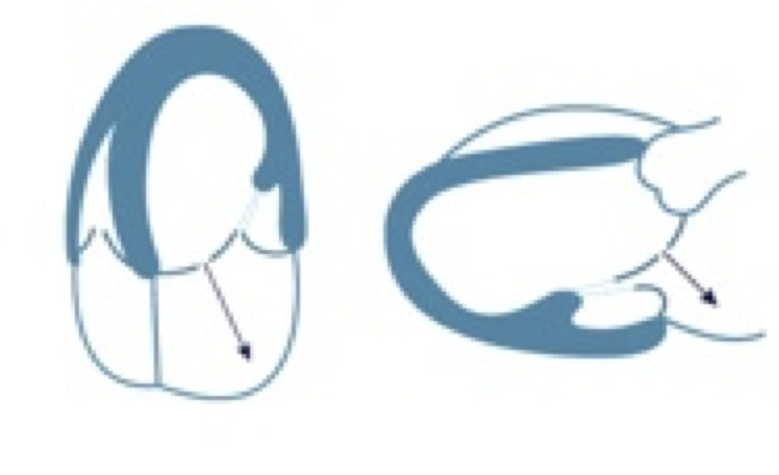
 
Perforation of the anterior mitral leaflet - left: 2D image of the four-chamber view. Note the thickening of the anterior leaflet (site of perforation, with and without color). Right: parasternal long-axis view: the color jet "goes through" the mitral valve.Commissural regurgitation
The direction of such jets may vary: it may either be directed towards the middle, or along the anterior or inferior left atrial wall. It is easy to miss the jets on "standard views". Their origin is best seen on a parasternal short-axis, or commissural view. Also use a five-chamber view (anterolateral commissure) and a coronary sinus view (posteromedial commissure) to detect regurgitation in these regions.

 
Mitral regurgitation with the origin of the jet at the posteromedial commissureAnnular dilatation
Annular dilatation causes a rather symmetric displacement of the leaflets. Thus, the direction of the jet will be central. It is broader on the two-chamber view than on the four-chamber view. In pure annular dilatation the origin of the jet is close to the annular plane.
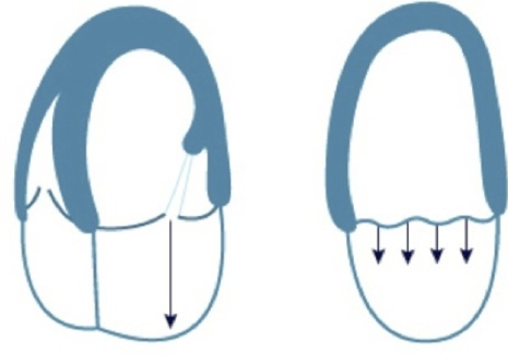

 
Annular dilatation with and without color Doppler. Note that the plan of closure is more towards the atria.Restricted physiology of both leaflets
The direction of the jet depends on the magnitude of restriction and its relationship with the anterior and posterior leaflet. When both leaflets are involved the jet will be central. In comparison to annular dilatation, in which the jet is also central, the origin of the jet will be farther within the ventricle. Restriction of leaflets is best seen on four- and three-chamber views.
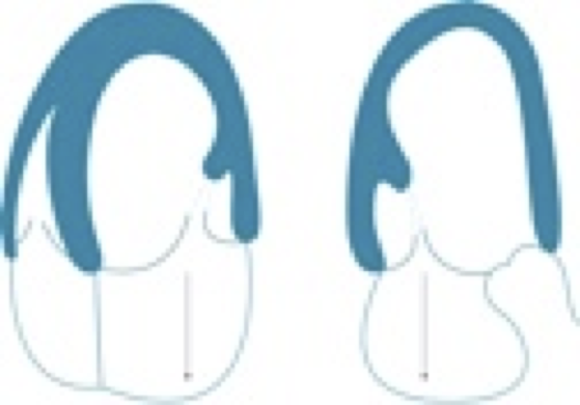
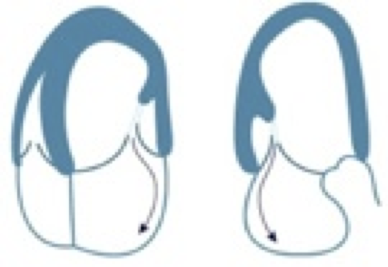
Restricted posterior leaflet
The origin of the jet is farther within the ventricle here. Due to the asymmetry of the valve, the direction of the jet is strictly posterior and lateral, following the angle of the restricted posterior leaflet. Use a three-chamber view to display the anterior, and a four-chamber view to display the medial component of the jet.
 
Restricted posterior leaflet with and without color Doppler. The posterior leaflet shows restricted motion and is pulled towards the apex.Mitral regurgitation in SAM
"Systolic anterior motion" pulls the anterior leaflet away from the posterior leaflet, but also distorts the posterior leaflet and causes an opening channel that directs the jet in posterior and lateral direction. Use color Doppler in a parasternal long axis, and a three-chamber view to visualize the direction of the jet.


Mitral regurgitation in a patient with hypertrophic CMP. The jet is directed more posteriorly.