Pleural Effusions and consolidations
November is Lung Cancer Awareness Month, and we want to emphasize the importance of early detection of lung cancer. Although lung cancer is not typically diagnosed using ultrasound, certain ultrasound findings, such as pleural effusion, can sometimes indicate an underlying malignancy. In this blog post, we have compiled a quick overview of pleural effusion and lung consolidations in relation to the risk of malignancy. In our comprehensive Lung Ultrasound MasterClass, we delve deeply into the interpretation of pleural effusions, lung consolidations, and other ultrasound findings and their associations with various comorbidities and systemic pathologies.
Pleural Effusions: Transudate vs. Exudate
Keep in mind: Pleural effusions and consolidations can often be directly or indirectly associated with malignancies—whether lung cancer itself or metastasis from other types of cancer.
When you detect a pleural effusion in patients, take a closer look at its characteristics on ultrasound:
Transudate: Typically appears echolucent (clear and watery content) on ultrasound and corresponds to conditions like heart failure or cirrhosis.
Exudate: Often contains echogenic content, septations, or fibers, reflecting a protein-rich, inflammatory nature. This type of effusion is commonly associated with malignancies, infections like pneumonia, or inflammatory conditions. While exudates can be classified in more detail, this general distinction is sufficient for now. After thoracentesis, laboratory analysis will provide detailed information about the composition of the fluid. Depending on your suspicion, analyses may include cell count and type (e.g., hematocrit), LDH, glucose, microbiology, and cytology, etc.

How does an exudate develop?
Exudates arise due to increased microvascular permeability or disrupted lymph drainage and are seen in infections or malignancies like bronchiocarcinoma.
Consolidations in the Pleural Region
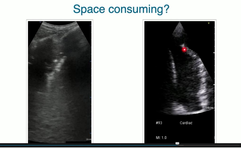
Another critical feature to monitor on ultrasound is consolidation. Here is an example of such a consolidation in the pleural region, showing typical features with a starry sky or solid-organ-like appearance. Consolidations may indicate malignancy, inflammation, or pneumonia. This case turned out to be pneumonia!
Ultrasound is, of course, not the primary diagnostic tool for this disease. CT and PET scans remain the standard for lung cancer diagnosis, staging, and treatment monitoring. However, lung ultrasound serves as a valuable complementary tool in specific contexts. It can be utilized to directly evaluate peripheral lung tumors, detect pleural effusions caused by primary tumors or metastases, and guide thoracentesis for detailed laboratory analysis of pleural fluid.
Learn more about lung ultrasound in our Lung Ultrasound MasterClass. Start your free lectures right away!


