Why Congenital Heart Defects Are No Longer Just a Concern for Specialists
Our Adult Congenital Heart Disease BachelorClass teaches essential congenital defects that every cardiologist should know, regardless of their work setting!
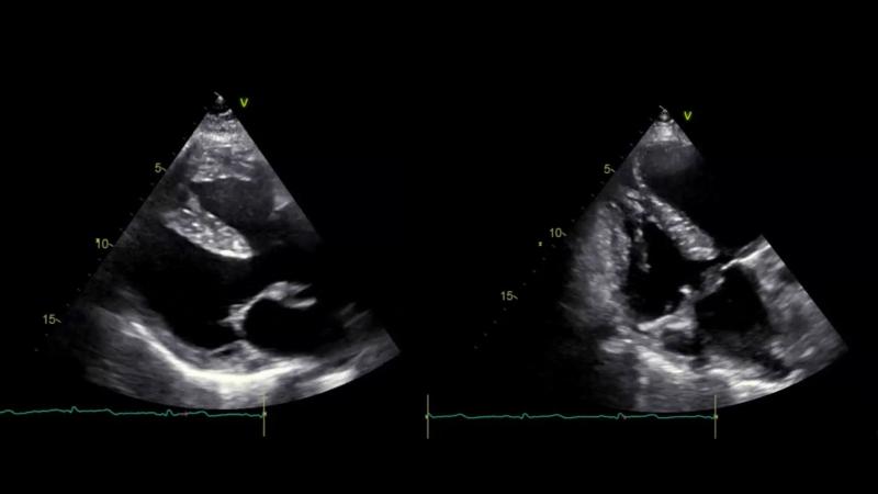
First, we are looking for a congenital cardiac disease characterized by four distinct anatomical abnormalities:
- Ventricular septal defect (VSD):
A “hole” between the right and left ventricles, leading to abnormal blood flow. - Pulmonary stenosis:
Narrowing of the pulmonary valve or pulmonary artery, obstructing blood flow to the lungs. - Right ventricular hypertrophy:
Thickening of the right ventricular wall due to increased workload. - Overriding aorta:
The aorta is positioned above the VSD, receiving blood from both ventricles.
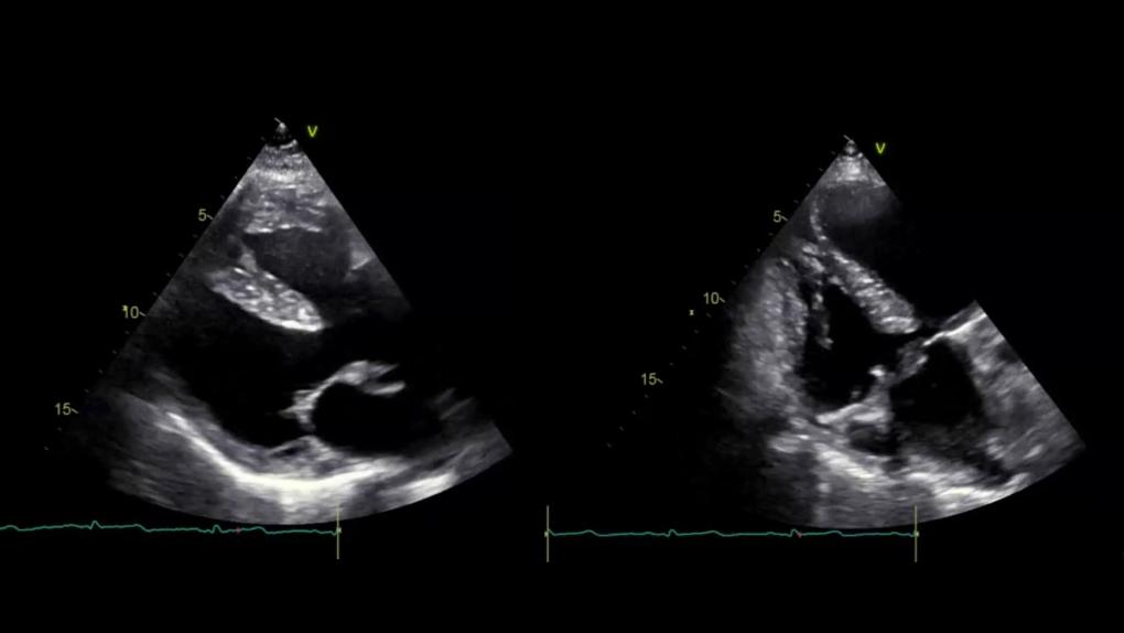
And here’s an interactive case showing (some of) its features. Can you answer all quiz questions? Click on the + in the video to interact!

Did you get it right? What is the name of the congenital heart defect that we are talking about? Find the answer to this case below and check if you are already well equipped or still need more training!
Adult Cardiologists Must Stay Up-to-Date on Congenital Heart Defects
Decades ago, infants with congenital heart defects (CHD) had poor prognoses. However, advancements in pediatric cardiology and improved medical care have allowed more of these patients to reach adulthood. Consequently, adult cardiologists are increasingly seeing patients with congenital heart disease (ACHD) in their practices. It is now essential for cardiologists and echocardiographers, who used to focus on acquired heart diseases, to be knowledgeable about managing repaired and unrepaired congenital defects and their sequels. Which is why we have created a BachelorClass focusing on just this topic – check out our free lectures below!
The Complexity of ACHD Management
Congenital heart defects are structural abnormalities present at birth, ranging from simple issues like atrial and ventricular septal defects to more complex conditions such as transposition of the great arteries and single-ventricle defects. Evaluating congenital heart disease (CHD) requires consideration of the initial anatomy, the surgical repairs performed, and the current physiological status. Each case demands careful evaluation due to the influence of these factors on the severity of the disease.
For you as a treating adult cardiologist, this means…
Recognising Residual and Repaired Lesions: Many ACHD patients have undergone surgical corrections but still require lifelong monitoring for residual defects, or arrhythmias, or heart failure.
Understanding Unique Hemodynamics: Unlike acquired heart disease, ACHD patients often have non-standard circulation patterns due to their congenital condition or previous surgeries. Some interpretations might be counterintuitive when applying your general cardiological knowledge to this patient group. For instance, high jet velocities in ventricular septal defects (VSD) are not a cause for concern; instead, they indicate that the VSD is very small and likely not hemodynamically significant.
Using additional echo views and techniques: To evaluate a wide range of congenital heart defects, it is essential to expand the variety of echocardiographic views that you master (like the supraclavicular view, the modified parasternal short axis view, etc…). A deep understanding of both anatomy and hemodynamics—encompassing both physiological and pathological states—is crucial. The echocardiography protocols in specialised clinics include the assessment of situs, atrioventricular connections, and ventriculoarterial connections, and importantly, integrate cardiac MRI.
What kinds of defects will you come across?
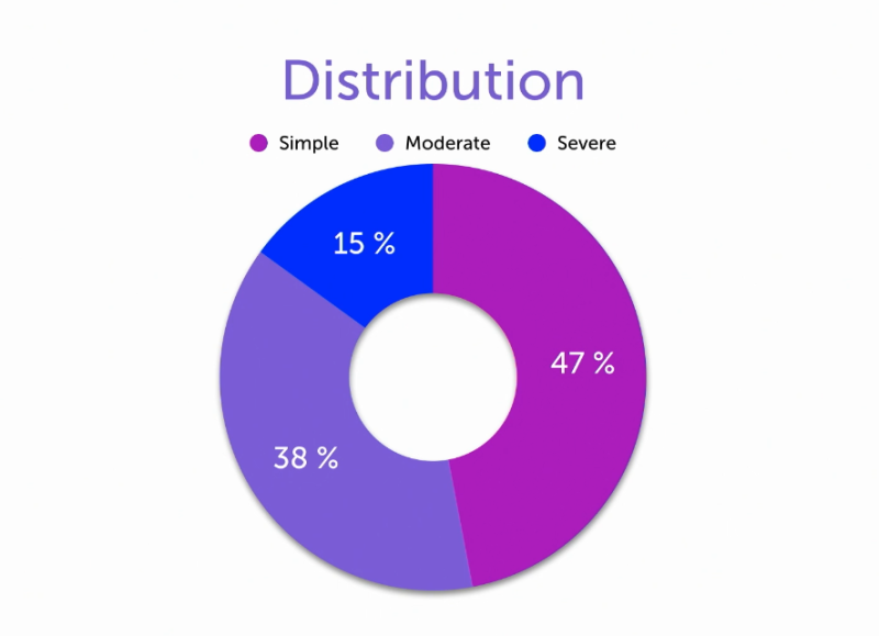
In most adult echocardiography labs, nearly 50% of patients have simple congenital heart defects (CHD). Specialized clinics may have a higher percentage of patients with moderate to severe defects, requiring a deeper knowledge of various surgical and nonsurgical management options. It’s also crucial to understand how these interventions affect patients' physiological conditions when they arrive in the echo lab.

The correct diagnosis of our interactive case is:
Tetralogy of Fallot (TOF)
Tetralogy of Fallot (TOF) is a congenital heart defect consisting of four key features: ventricular septal defect (VSD), right ventricular outflow tract (RVOT) obstruction (often pulmonary stenosis), overriding aorta, and right ventricular hypertrophy (RVH). Cyanosis depends on the severity of RVOT obstruction and the resulting shunt hemodynamics, specifically the degree of right-to-left shunting across the VSD. Greater obstruction leads to more deoxygenated blood bypassing the lungs and entering systemic circulation. Post-repair complications include right ventricular dysfunction, arrhythmias, and pulmonary valve regurgitation.
TOF requires lifelong surveillance!
Want to learn more? Check out our Adult Congenital Heart Disease BachelorClass – it is now available with a 40% discount!


